What is the nuclear picture?
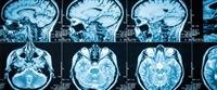
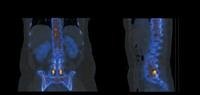
The answer to a question is what is the nuclear image in that it is a mechanism for imaging the different parts of the human body, by injecting a radioactive material into the body through the vein, or by eating the radioactive material through the mouth in order to perform some tests, and in this picture a body The human being is the radioactive source, and the imaging device captures the radiation emitted from the body, and it is converted by computer to a digital image, then it is sent to a nuclear medicine specialist in order to read the image and diagnose the disease, and this imaging mechanism differs from other radiology devices that are used from In order to evaluate the functions of the body''s organs, while the other x-rays perform an anatomical evaluation of the organ, that is, what the organ looks like, and also in this image, the patient is the source of the rays and not the device as in other types of radiographs.
Cases where the nuclear picture is used
After knowing what a nuclear image is, the types of nuclear tests will be mentioned, as each examination employs a specific technique and radioactive material and its own procedures, and the nuclear picture consists of three steps that begin by injecting the radioactive material into the body, then the required images are taken and then the images are evaluated in order to Diagnosis, the injection of radioactive material and imaging may take from several minutes to days, depending on the tissue that is imaged and on the radioactive material used, and in the cases where the following nuclear image is used:
Kidney examination: This is to assess any problems in it, including functional problems with the kidneys or blockage of blood flow.
Thyroid screening: which is used to assess thyroid function or to get a better evaluation of thyroid nodules or the masses in them.
Bone examination: The nuclear image helps to diagnose any degenerative changes or inflammation in the joints, and helps in finding bone diseases and tumors, and helps in determining the cause of bone pain or inflammation.
Gallium examination: which aims to diagnose active infectious diseases, inflammatory diseases, tumors, or abscesses.
Cardiac examination: The nuclear image can be used to check any problem with blood flowing inside the heart, and the picture also determines the extent of damage to the heart muscle after a heart attack, and also enables the measurement of heart functions.
Brain scan: that helps check for any brain or circulatory problems.
Breast examination: A nuclear breast examination is performed in addition to a mammogram, in order to locate the cancerous tissue.
The risks and complications of the nuclear picture
After defining what a nuclear image is and its use, it is worth mentioning the risks of the nuclear picture and its complications, as the risks of the nuclear image are exposure to a large amount of radiation, which may lead to damage to tissues and organs or the body or may increase the risk of developing cancer, but in general the amount of radiation Which is used for diagnosis in nuclear imaging is few and similar to the dose of radiation used in axial tomography and x-rays, and therefore the nuclear image is relatively safe, especially since the benefits obtained from the image outweigh the risks and that the radioactive material used in the nuclear image is not considered a colored dye or a drug and therefore It does not cause any side effects