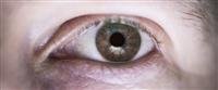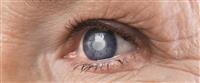

Glaucoma damage to the optic nerve This is a confirmed cause of blindness. It usually occurs when fluid pressure inside the eye increases, causing damage to the optic nerve. No symptoms appear at first, but a comprehensive examination of the eye can reveal this cause. People at risk should do tests
Periodic at least every two years. These tests include
Persons over the age of sixty
People whose families develop glaucoma
Early treatment can help protect the eyes from vision loss, and includes the use of eye s and / or surgery.
an introduction
Glaucoma is an eye disease that can end up blind if not treated.
Millions of people suffer from open-angle glaucoma, the most common form of glaucoma. Half of these patients are unaware that they have this disease.
This tutorial provides information about glaucoma and how it can be treated.
Eye anatomy
The eye is the organ of vision, which is a very complex organ that collects light and focuses it on the posterior wall of the eye in a way that allows the patient to see.
The cornea is the transparent front part of the eye. It allows light to pass into the eye. The rest of the eye is covered with an outer layer called the hard layer.
An additional layer called conjunctiva covers the front of the eye.
The light shines on the iris, the colored part of the eye. The hole in the center of the iris is called the pupil or pupil. The iris adjusts the amount of light entering the eye by changing the pupil''s amplitude.
After light passes from the pupil or pupil, the light passes through a transparent lens that focuses the light on the bottom of the eye, and is thus very similar to the viewfinder.
The wallet holds the lens in place.
Light rays pass through a transparent substance called the vitreous after passing through the lens and before it reaches the back wall of the eye.
The bottom of the eye is called the retina. The retina converts the traffic signals into electrical signals.
Electrical signals pass through the optic nerve to the brain, which translates it into human-seen images.
The eye is a room or a front room, and a room or a back room. The anterior chamber is located in front of the iris and the posterior chamber is located behind the iris and in front of the lens.
The transparent fluid that fills the anterior chamber forms between the iris and the lens, and is then absorbed via a sponge network located in an area called the angle.
If the fluid absorption velocity is less than the speed at which it forms, it accumulates in the anterior chamber. When the fluid accumulates, the pressure in the eye increases and the tension in it increases.
This increase in pressure causes eye and optic nerve damage and can lead to blindness.
Types of glaucoma
There are five types of glaucoma
Open angle glaucoma
Low pressure glaucoma or normal pressure glaucoma
Closed-angle glaucoma
Congenital glaucoma
Secondary glaucoma
Open angle glaucoma is the most common form of glaucoma. Eye pressure rises over time at a relatively slow pace. This glaucoma can usually be treated with medication or surgery.
Low pressure glaucoma or normal pressure glaucoma is treated in the same way as open-angle glaucoma treatment. But the symptoms here do not occur as slowly as in open-angle glaucoma, they may occur suddenly.
Closed-angle glaucoma is an emergency. This type generates a sudden increase in intraocular pressure. Signs of severe pain, nausea, red eye, and blurred vision.
If closed-angle glaucoma is not treated, blindness can occur within a day or two. Usually, ambulatory laser surgery can open the blockage and save vision.
Congenital glaucoma affects children born with ocular deformities that hinder the normal drainage of fluid. Its signs are cloudy eyes, intense light sensitivity and frequent tears.
Usually surgery is the best treatment, and there is an excellent opportunity for the child to recover and enjoy a good vision.
Secondary glaucoma is a complication of another disease. Treatment may be medicinal or surgical.
The diseases that can lead to secondary glaucoma are
Eye surgery
Advanced cataract
Eye injuries
Some tumors of the eye
Ocular uveitis
Diabetes
Take cortisone medications
Symptoms of glaucoma
Glaucoma may be asymptomatic in its early stages.
If patients with glaucoma are not treated, they may suddenly find that they are unable to side or peripheral vision. They may feel as if they are looking through a tunnel. This condition is called tunnel vision.
Over time, the glaucoma patient''s vision may increase until he becomes completely blind.
Symptoms of glaucoma include eye pain and headache.
Glaucoma usually affects only people over the age of 60 Except for congenital glaucoma, but it is a rare condition.
Some races have this disease more, for example, African Americans over the age of forty are at greater risk of developing glaucoma. But the reasons for this are still unknown.
A person is more likely to develop glaucoma if any of the following conditions is met
The presence of a glaucoma injury in the family
The presence of other ocular diseases
The presence of previous surgeries on the eye
Diabetes
Diagnosis of glaucoma
Early diagnosis of glaucoma is important to avoid blindness and blindness.
Most patients do not feel the symptoms immediately. Glaucoma can only be detected through regular periodic eye examination.
A close eye examination includes a test
Optical power measurement
Drawing field of view
Examination of the fundus of the eye after dilating the pupil
Eye pressure measurement
Measurement of corneal thickness
In measuring visual ability, the doctor asks the patient to shapes in the dashboard, to measure the eye''s ability to see it from different distances.
As for the field of vision test, it measures the patient''s side or peripheral vision. It helps the doctor to know if the patient has a lack of peripheral vision, because this is evidence of the presence of glaucoma.
Mydriasis allows the doctor to see the optic nerve more clearly, to check for signs of damage.
The doctor applies eye s to dilate the pupil. After examination, the near vision remains blurred for several hours.
Eye pressure measurement determines the amount of fluid pressure within the eye. There are several types of intraocular pressure gauge.
One species uses a purple red light to measure pressure. There is an air pressure measurement by eye pressure, which measures eye resistance to airjet.
While measuring the thickness of the cornea, the eye is numbed with a and an ultrasound probe is placed to measure the thickness of the cornea.
Treating glaucoma
After diagnosis, glaucoma can be treated in several ways.
Initially the doctor tries to use the medicine. If the medication does not help to control eye pressure, surgery may be required.
It is sometimes advisable to have laser surgery at first.
Glaucoma medications are in the form of eye s and oral tablets. Some of these medications reduce fluid formation. Others work to reduce intraocular pressure by facilitating the drainage of fluid from them.
Glaucoma medications are usually taken several times a day.
Most people do not have problems caused by these medications, but some patients experience a headache or side effects on other parts of the body. Drops may cause tingling or burning sensation in the eye, and they may also cause redness.
If the doctor decides to give medication to a glaucoma patient, he will explain to him how to use it. Before starting treatment for glaucoma, the patient must tell the ophthalmologist about other medications he is taking.
There are many medications for glaucoma. If the patient has a problem using one of the medications, he must inform the doctor. It may be appropriate for him to use another medicine, and the doctor may limit himself to adjusting the dose.
It is important for the patient to use the s and tablets as long as they help reduce eye pressure. Sometimes the patient tends to stop taking the drug because glaucoma rarely gives clear signs.
Glaucoma surgery
If the medications alone are unable to maintain low eye pressure, the patient may need laser surgery, which is called "trabeculoplasty". More often than not, the patient still needs glaucoma medications even after surgery.
The eye is anesthetized with special s. The patient sits opposite the laser, and the doctor places a special lens on the eye.
A high-energy light beam is directed onto the special lens and reflected onto a special network to reach the inside of the eye. The patient may see flashes of bright red or green light.
The laser produces 50-100 burns, separated by equal distances. These burns extend the drainage pores of the network.
Non-laser surgery may be required. The goal of non-laser surgery is to a new opening for fluid drainage.
A small piece of tissue is removed from the white of the eye called "sclera", which opens up a new field for drainage of fluid from the eye.
Surgery does not leave an open hole in the eye. There is a thin, transparent tissue covering the whites of the eye called the conjunctiva. The fluid flows through the new opening under the conjunctiva, and is drained from the eye.
Conclusion
Glaucoma is a serious eye disease that can cause blindness if not treated.
The best way to diagnose glaucoma is by regular periodic eye exams. Every African American who is over forty and anyone else over the age of 60 must undergo regular eye examinations.
Early diagnosis and treatment can save vision in most patients so they can continue enjoying their lives!