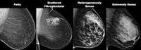

Digital mammograms, television imaging and magnetic resonance imaging, most notably EliteDoctorOnline Jeddah: «Middle East» According to the standards of the American Cancer Society, early detection of breast cancer is done by mammography (mammography), for women aged 40 years and over, annually. Then a breast examination is clinically performed by a specialist, every three years in the twenties and thirties, and annually at the age of forty and over. And by self-examination of the breast, starting from the age of 20 years, monthly, after learning the correct method by a doctor or nurse. Thus, the girl or lady is aware of the nature of her breast and thus it is easy for her to know any changes that occur to it, and to consult a doctor immediately.
Recent developments, Dr. Iman Baroum, head of the radiology department and consultant of diagnostic radiology at King Abdulaziz Hospital and Oncology Center in Jeddah and vice-president of the organizing committee for the activities of the Breast Cancer Awareness Week in Jeddah, indicated that there are recent developments that have entered the field of mammography, such as: a digital mammogram: in which the x-ray film is replaced by sensors Or probes that convert x-rays into electrical signals that give the final image of the breast that appears on the computer screen connected to the device. CAD programmer: to discover abnormal areas in the mammogram, such as tumors and calcifications, which helps in drawing the attention of the radiologist while making the report to these changing areas for more accurate consideration and analysis. The mammogram is used to perform a "periodic scan" of the breast, without the presence of symptoms, and it helps in early detection of cancer even two years before the patient or the doctor senses it. The mammogram is also used to "diagnose" in the event of a complaint in the breast or the presence of abnormal signs in the mammogram.
Imaging procedure, and Dr. Iman Baroum adds that the mammogram has preparations, including
It is preferable to do it a week after the menstrual period, as the breasts are sore before and during the period.
Inform the doctor about any new symptoms in the breast, any previous operations, any use of hormones, and any family or personal history of breast cancer.
Informing the radiologist of any previous breast examinations to make a comparison, it is very important.
Inform the radiologist, before the examination, of any symptoms or problems in the breast and about any possibility of pregnancy. - Do not apply deodorant, powder or cream in the armpits or breast area on the day of the examination, because they appear on the image such as calcifications.
And Dr. Imanbarum adds that the work of the mammogram does not require hypnosis, and the examination takes about 20 minutes for the breasts. The breast is fixed, by the radiologist, in the required position in the device to take two views of each breast, the first from top to bottom and the second in the position of the breast tilted at an angle of 45 degrees. Then a gradual pressure is applied to the breast for the following reasons:
Straightening the breast tissue in a homogeneous way so that no tumors or calcifications are covered by the breast tissue.
The use of a radiation dose is lower because the thickness of the breast becomes less.
Breast stabilization because less movement during imaging leads to vibration in the image, which may lead to not seeing the tumor or calcifications, even if they are present, and may also ask her not to breathe for seconds.
Advantages and shortcomings of the advantages of the mammogram. Diagnosing tumors in a very early stage, especially with the mammogram, where the treatment is complete and final, and there are no harm to radiation due to the small radiation dose used. Despite that, it has some shortcomings and shortcomings, such as:
False positive results (from 5-15%), requiring other tests such as taking a sample. Difficulty distinguishing definitively, in some cases, between benign and malignant tumors and calcifications, and other examinations are then performed.
Not all cases of tumors can be seen on a mammogram.
Aesthetic breast enlargements, the presence of which may hinder the vision of tumors. And if there is a suspicious area when reading the mammogram, the radiologist will do:
Additional mammograms.
Television imaging of the breast to find out if this area is a cyst or a solid tumor.
Make a breast magnetic resonance.
Taking a sample, under local anesthesia, from the tumor or the area of calcifications and examining it microscopically in a tissue laboratory to see whether it is benign or malignant (80% of the samples are benign).
Television mammography, television mammography is used as a primary procedure for examining the breast in women under 35 years of age and pregnant women, to re-examine a suspicious area in the mammogram to see if it is a cyst or a solid tumor, whether it is palpable or not, and to examine the breast that appears thick on the mammogram, as well as when taking Breast samples.
Magnetic resonance of the breast (MRI) is performed between day 5-15 of the menstrual cycle, and a strong magnetic field and frequency waves are used to take pictures in several directions (there are no rays at all), and crackling sounds are heard during the examination as a result of the MRI, an intravenous dye is taken during the examination to show any changes more clearly The examination usually takes 30 - 60 minutes. This test is used to:
Diagnosis - Diagnosis of breast cancer cases that are difficult to diagnose with a mammogram.
The best examination for examining the enlarged breasts beautifully, either to examine the cosmetic cyst or to detect cancer in the surrounding breast tissue.
Determining the degree of spread of cancer in the breast, which determines the appropriate type of treatment, either surgically, chemically, or otherwise.
Breast follow-up after primary cancer treatment.
Second: The survey (under study) of women under the age of forty who have a high susceptibility to breast cancer, such as having a strong family history of breast cancer (mother, sister, aunt, and aunt), and the presence of cancer genes such as BRCA1 / BRCA2.
Benefits of magnetic resonance
It is very sensitive to detect even small changes and diagnose dense breast tissue.
Imaging of breast magnifiers and diagnosing any leakage.
Examination of an inverted nipple to see if the cause is cancer.
To assess the extent of cancer in the breast.
Determining the type of operation for breast cancer, is it the removal of the tumor only or the whole breast?
Breast examination after treatment to find out if there is a lump remaining or if the disease has returned after the operation.
Breast examination to find out if there is a lump in cases of enlarged axillary lymph nodes as a first symptom.
It can be used for screening in women under the age of forty, who have a high risk of breast cancer.
It is taken on the MRI of the breast that it is very expensive, and it takes 30-60 minutes compared to a mammogram (10-20 minutes), and it needs to use an intravenous dye, and finally the patient should be able to tolerate narrow places and not be afraid of them (claustrophobia).