
by removing a brain stem tumor
In a unique global achievement, the team of the Neuroscience Center at King Fahd Medical City succeeded in surgically removing one of the types of brain stem tumors. The traditional treatment for this type of tumor was radiotherapy, according to the testimony of more than one specialized center worldwide.
The child Abdullah was referred to King Fahd Medical City from a secondary care hospital, and he suffers from symptoms of high brain pressure, and the condition was diagnosed as a brain stem tumor requiring radiotherapy under the supervision of a specialized pediatric neurosurgeon in King Fahd Medical City, and the city’s examinations proved The presence of a tumor inside the middle part of the brain stem called (Pones), with a large part of it extending to the front part of the brain stem, which is difficult to access surgically due to the presence of dense parts of the bone of the base of the skull in front and on both sides of the middle part of the brain stem, and the case was discussed in detail and accordingly it was agreed By starting with taking local and international opinions about the possibility of surgically removing the tumor, which would enable the child not to expose the child to radiotherapy, whose effects are known in children’s age.
The Associate Executive Director of Medical Departments, Director of the Neuroscience Center, Skull Fund and Cerebral Vascular Surgeon, Dr. Mahmoud Al-Yamani explained during the press conference held yesterday in the Medical City to highlight this process that all the references that were consulted locally and in the United States and Canada reported that the tumor could not be eradicated. Because it is located in an area difficult to enter first and also because there is a risk of removing part of the tumor that lies within the middle part of the brain, as the area of the brain stem is very narrow and contains very sensitive areas such as the area of consciousness, the area of the nerves of the face, hearing, swallowing, breathing, heartbeat and other Important vital functions.
He added that the problem was analyzed on three axes by accessing the front of the brainstem, which requires removing a large part of the skull base bone from the right side, as well as consulting specialists to monitor nerve functions during operations and under anesthesia, and they reported that it is possible to monitor the part in which the operation will take place during the operation. And before part of the tumor is removed, to ensure that there are no vital parts inside the tumor, as well as the possibility of magnetic resonance imaging during the operation to ensure that none of the sensitive parts of the brain stem are damaged and that the excision remains within the boundaries of the tumor, indicating that with the completion of the main axes that were made He mentioned it, which is not available in any other hospital in the Middle East, which is the magnetic resonance device inside the operating room, monitoring the nervous system during operations with the presence of the magnetic resonance device in the operating room, and the possibility of accessing the front part of the middle of the brain stem by removing part of the bone in the base of the skull underneath Neural planning, the team concluded that the success rate will be high.
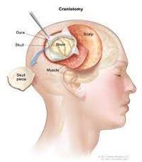
He indicated that the subject was discussed in detail with the patient''s parents, and after the parents were convinced of the surgical procedure and the potential for harm and benefit from it, the operation was planned in two stages. In the first part, large parts of the skull base bone were removed to reach the front of the middle part of the brain stem, and it took eight continuous hours. In it, the use of the latest techniques of skull base surgery and access to the area to be accessed, and the second operation took place in the brain surgery suite equipped with an intraoperative magnetic resonance device, a tumor locator and nerve monitoring devices during the operation, and it took 14 hours to enter the front of the middle part of the brain stem and remove the tumor The developing from the middle part of the brain stem extending to its front, and a large part of the inside of the tumor was removed and microscopic examination of the cells and the pathology department reported that the tumor is of a special type of astrocytoma called Pielocytic Astrocytoma, which reassured the team that the remaining part of the tumor latent in the center of the brain stem and which is not More than 20% of the entire tumor does not need to be removed. This is because the removal is dangerous, and because this type of tumor does not grow after removing part of it and in Sometimes the remaining part becomes atrophied after parts of the tumor are removed.
He added that the team returned to search in modern and old scientific references for any similar case in which the front of the brain stem of a child was reached to remove a tumor of this type (almost complete resection), but it was not found, because these tumors either grow inside the brain stem and these are treated radiographically. And the other type grows to the back of the brain, and these are removed by traditional methods. The distinguishing feature in this case is that the tumor from the middle part of the brain stem extends to the front part of the brainstem in the posterior cranium of the skull, which made the removal by normal methods impossible.
It is noteworthy that the patient is in excellent condition and is preparing to leave the hospital in the coming days, and the medical team participating in the operation, in addition to Dr. Mahmoud Yamani, consists of a consultant pediatric neurosurgery who supervises the training program for neurosurgery of the Saudi Commission for Health Specialties Dr. Abdul Rahman Sabbagh, who is the consultant supervising the case. Dr. Reem Al-Bunyan, Head of the Department of Neurophysiology (Nerve Planning and Monitoring) - Acting Head of the Neurology Department, Dr.Ahmed Abdel Moati, Consultant Pediatric Neurology - Consultant Pediatric Neuropathy and Nerve Monitoring During Operations and Supervisor of the Training Program for Pediatric Neurology, Dr. Ayman Al-Bunyan is a consultant pediatric neurosurgeon, Dr. Muhammad Al-Takrouri, a consultant anesthesia for brain and nerve operations, in addition to the medical team consisting of neuro-monitoring and planning technicians, assistant physicians, residents of neurosurgery, anesthesiology and medical imaging technicians.