
Ultrasound examination is one of the best safe ways to detect the health of the fetus Ultrasound scanning technology is one of the best safe ways to detect the health of the fetus and ensure that it is free from birth defects that about 2-4% of live births suffer from various reasons such as genetic diseases or defects Chromosomes, which constitute 15-20%, in addition to other causes such as mother''s diseases such as blood sugar, taking certain medications during pregnancy, or infections. While 65-75% of them are of unknown cause, it is recommended to do an ultrasound examination for all pregnant women to avoid many of the risks that may occur to the mother or fetus during pregnancy.
It is also to ensure the health of the pregnancy and that it is free from some complications such as placenta previa, causes of bleeding and multiple pregnancy, noting that these complications require careful follow-up and may require delivery in a specialized hospital and some of them may require a cesarean delivery.
There are some special cases in which the fetus is treated before birth by transferring blood to the fetus through the umbilical cord for cases of fetal anemia or by injecting treatment into the amniotic fluid for cases of thyroid gland disorders of the fetus and its enlargement or giving the mother the medicine and then it reaches the fetus through the placenta in cases of acceleration Fetal heartbeat.
Ultrasound is performed in 4 different stages during pregnancy
The first stage: determining the age and safety of the pregnancy
Between 6 and 10 weeks and is performed: the importance of this stage in the ultrasound examination is:
Diagnosis of the presence of a fetus inside the uterus and fetal pulse
Determine the age of the fetus
Diagnosing a twin pregnancy
Examination time: 5-10 minutes
The second stage: the first examination of the safety of the fetus
Between week 11 and week 14, when the NT method is used, this examination is very important because it provides initial and early information about the health of the fetus and the volume of fluid under the skin of the back of the neck of the fetus is measured, as an increase in this size may indicate the presence of a chromosomal defect such as Down syndrome. Or to the presence of a congenital defect (congenital heart disease) or to the presence of other defects.
Duration of the examination: between 5 and 15 minutes.
The third stage: a detailed examination of the fetus''s organs
Between week 18 and week 22 at this stage we need to perform two types of ultrasound
The first type: the detailed examination is a thorough examination of the fetus’s organs to ensure the safety of the fetus and the absence of basic congenital defects that result in certain procedures that help us to early diagnosis and treatment of some of these ailments during pregnancy or to arrange the necessary procedures before birth for the safety of the fetus, which includes determining the appropriate time and place for childbirth and providing Medical staff treating these cases.
During this period, the cervical length is checked to detect the possibility of premature labor.
Examination time: 20 to 40 minutes
Type II: detailed examination of the fetus''s heart
It is an accurate examination in which the performance of the heart, valves, arteries and veins connected to the heart is examined and the presence of a large hole in the septum between the right and left ventricle is excluded.
Note that early diagnosis of these congenital heart defects is of great importance, because some of them require birth in hospitals that have special expertise and equipment for diagnosis and treatment in the first hours or days after birth.
Examination time: 20 to 40 minutes
Fourth stage: examination of the growth and health of the fetus
Between the 23rd week until the end of pregnancy
At this stage, the fetus’s growth is confirmed as required, the amount of amniotic fluid surrounding the fetus is examined, the placenta and the fetus are placed.
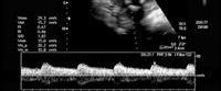
In diagnosing delayed fetal growth, Doppler and fetal monitoring are used
The blood flow in the umbilical cord, arteries and veins of the fetus, the amount of amniotic fluid surrounding the fetus and the fetal breathing movement are examined to determine the best time for delivery.
Examination time: 20 minutes
Safety ultrasound examination
Ultrasound is safe for you and your fetus, because it does not contain harmful ionizing radiation such as X-rays
In the end, we wish you a safe pregnancy, and remember that your fetus has tests that must be adhered to for the sake of its safety