
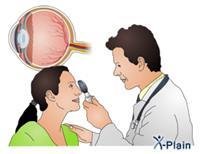
Flashes and floaters in the field of vision are common, and do not usually indicate a serious medical condition. Floaters appear as small spots, black spots, circles, lines, spiderwebs, or any other shape in the field of vision. They usually appear relatively transparent gray or white. The floaters may remain in one place, and they may move rapidly out of the field of vision. It is of little medical importance, unless it affects vision. As for the flashes, they appear as flashes of light, and their appearance is sometimes accompanied by floaters. Flashes that last ten to twenty minutes may accompany a "migraine" crack. Flashes caused by the vitreous gel separating the eye from the retina are a sign of aging and should disappear within a few weeks or months. There aren''t any medications or eye s that make the floaters disappear, but the brain usually ignores them. If the flashes significantly affect vision and do not disappear over time, your doctor may recommend surgery to remove the vitreous gel from the eye and replace it with a saline solution. If the floaters appear suddenly, or their number increases rapidly, the patient should see an ophthalmologist immediately because this may be an indication of a serious eye problem that needs urgent surgical intervention.
an introduction
Sometimes people see moving spots or sparks in their field of vision. And they call these sensations flying objects or flying flies or smudges.
Almost seven out of ten people see UFOs at some point in their lives. Sometimes a person next to these objects sees a flash of light.
Seeing flying objects and flashing is very common and is not usually a sign of a serious health problem. But a sudden onset of it may indicate a serious eye problem such as a retinal tear or a retinal detachment.
This tutorial explains the meaning of flying objects and flash, as well as their symptoms and causes, and when they are indications of a serious illness. The program also deals with diagnosing and treating flying objects, flashing, and caring for each person himself.
Anatomical overview of the eye
Knowing the parts of the eye has something to do with talking about flying objects and flashes. Especially the anatomy of the eye.
Light falls on the cornea of the eye at first. The cornea is the transparent covering located at the front of the eye.
After that, the light travels to the bottom of the eye, passing through the pupil or pupil. The pupil is an opening in the center of the colored part of the eye. We call the colored part the iris.
The iris controls the amount of light entering the eye by controlling the size of the pupil or pupil.
After light passes through the pupil or pupil, it reaches a transparent lens that focuses light rays on the back wall of the eye. This lens performs the role that the camera or camera lens does.
After the focused light rays pass through the lens, they travel through a transparent gel called the "vitreous" and finally fall to the base of the eye where the retina is located.
The retina converts light into electrical signals that travel through the optic nerve to the brain, which translates them into visual images.
The middle part of the retina is called the "macula" and it allows people to see things clearly in front of them.
The rest of the retina is called the periphery, and it allows people to see things on both sides. This vision is called "peripheral vision" or "side vision."
The retina, like all organs in the body, needs blood to function. And the retina is supplied with blood through tiny blood vessels.
Symptoms
People with UFOs see moving shapes within their field of vision.
Flying objects can appear as spots, circles, lines, or clouds, spider web shapes, or other shapes. It usually appears gray or white, and is sometimes slightly transparent. They can appear moving or stationary.
Roughly seventy percent of people see UFOs. The easiest way to see flying objects is to look at a flat background, such as looking at a white wall or at a blue sky. A person can close one of his eyes and look at the white space with the other eye. If he sees a moving spot or a moving shape, then these things are flying objects!
Flying objects are usually small and quickly disappear from the field of view. Therefore, it has no medical significance unless it becomes large and causes blurred vision.
Most importantly, flying objects can be an indication of serious eye problems such as retinal detachment that can lead to severe vision impairment and blindness.
If flying objects appear suddenly or if their number increases rapidly, she should see an ophthalmologist immediately, as he may have a serious eye problem.
Sometimes a flash of light may appear with flying objects. This flashes like flashing lights or light strips, although there are no bright lights around the patient. This flash is like the feeling of "seeing the stars" when a person is hit on the head. It usually appears at night or in dark places.
To some people, the blinking appears as chipped lines or "heat waves" in both eyes. The blinking usually lasts from ten minutes to twenty minutes. This type of blinking is usually caused by the spasm of some blood vessels in the brain, which is called "migraine". A headache sometimes follows the sister.
the reasons
The vitreous that fills the eye is composed mainly of water, but it also contains proteins and other molecules. Focused light rays pass through the vitreous to the retina.
If any object is found between the rays of light and the retina, its shadow falls on the retina. Thus we conclude that UFOs are shadows caused by objects suspended in the vitreous. When we see flying objects, this actually means that we see shadows falling on the retina.
Where do these objects or objects in the vitreous come from? Flying bodies come from the vitreous itself or from cells of the tissue surrounding the vitreous.
Protein fibers can clump in the vitreous, and these clumps can block the path of light and cast a shadow on the retina. This type of flying object is usually small and may appear in the form of a spider web, transparent circles, or small worms, and their appearance is not accompanied by any flash. It remains in the vitreous permanently and one gets used to ignoring it.
Sometimes blood vessels in the retina can bleed if you undergo a vitreous lift. Generally, the red blood cells that result from bleeding appear as smooth black spots, or they may appear like a swarm of mosquitoes or a cloud of smoke. The flying bodies caused by the red blood cells usually disappear after the blood is absorbed. But it could take several months for it to disappear completely.
With age, the vitreous becomes dense and begins to shrink, separating slightly from the retina. This separation is called posterior vitreous detachment. The fragments of the particles resulting from this detachment penetrate the vitreous and form flying bodies.
Typically large-sized plane bodies appear when posterior vitreous detachment occurs. This type of flying object can look like a spider web or like a curtain blocking part of the field of view. Posterior separation of the vitreous causes the vitreous to exert a pulling force on the retina, causing the flash to appear. When a patient sees a flash and a large number of flying objects this is most likely a sign of posterior separation of the vitreous.
Posterior vitreous detachment is more likely if the person has previously undergone cataract surgery or if he has had eye trauma or an intraocular infection. Also, the occurrence of posterior separation of the vitreous in people with short vision, more than in people who have normal vision.
Most of the time, the vitreous separates cleanly from the retina without causing any other problems. The flash gradually decreases until it completely disappears. Flying bodies that result from posterior separation of the vitreous may persist, but become less bothersome within two to three months.
In some people, detachment of the vitreous can cause a tear in the retina. This tear can cause the retina to separate from the back wall of the eyeball, which is a serious condition that may lead to blindness. It is a complication that affects the retina.
Retinal complications
If the vitreous is very attached to the retina, or if the retina is weak in one place, it is possible that a rupture of the retina occurs during the separation of the vitreous from it.
After a retinal tear, there is a high chance of vitreous fluid leaking out through the site of the tear. This causes the retina to detach from the back wall of the eyeball. This condition is known as "retinal detachment." And when the retina detaches, the patient may become partial or complete blindness in the affected eye.
Symptoms of retinal detachment are
A sudden increase in the number of flying objects
Light flashes
Vision impairment
Shade or curtain over peripheral vision. The shadow usually moves within hours, days, or weeks, moving into the central vision area
When the retina detaches, it becomes distant from the tissue that holds and nourishes it. The condition of the retinal cells deteriorates due to the lack of nutrition. If the retinal detachment extends to the central area of vision (the macula), the patient''s vision begins to decline.
The longer the central area of the retina detaches, the more severe the degradation of cells and the weaker vision. Therefore, it is advisable to diagnose retinal detachment and correct the situation before the detachment reaches the central vision area.
Even when retinal detachment reaches the central macula, repairing this detachment remains important to prevent complete blindness or to restore some of the lost vision. Therefore, the patient should see a doctor who specializes in eye surgery immediately in order to give himself the best opportunity to diagnose and treat retinal detachment before it reaches the macula.
The diagnosis
The doctor examines the patient''s eyes to see if what he sees of flying objects and flashes is caused by a tear in the retina or other eye problems. The ophthalmologist is the doctor who specializes in diseases and eye surgery.
The doctor will ask his patient to prescribe flying objects and flashes.
Then the doctor examines the patient''s eyes for any injury to the retina. The doctor cannot see the flying objects that the patient is seeing unless they are very large.
A doctor can see a small portion of the retina through the pupil. After dilating the pupil with special eye s, the doctor is able to see the entire retina with an instrument called a fundoscope.
Mydriasis is a very safe procedure. Most people experience only some blurred vision and increased sensitivity to light for several hours after mydriasis. During these hours, the patient may not be able to do any delicate work, such as reading or sewing.
The doctor asks his patient to look in different directions when examining his eyes. And he must strictly implement the doctor''s instructions so that he can conduct a comprehensive eye examination.
The doctor will apply gentle pressure to the patient’s eye with a stick of cotton on it or with a special, non-cutting metal instrument called a "sclera compressor." The pressure on the eye in this way, which is called "sclerotomy", allows the doctor to see areas of the retina that he cannot see otherwise, and is a necessary procedure for a comprehensive examination of the retina. This pressure may cause some discomfort to the patient, but this discomfort can be minimized by using anesthetic eye s.
The doctor uses a bright light to examine the retina. You will notice that everything appears to him to be a dark purple for a minute or so after the eye examination. After that the color gradually turns pink. This bright light is harmless to the eyes. In ten minutes or less, things should be looking normal again. If this sensation lasts longer than that, he sho