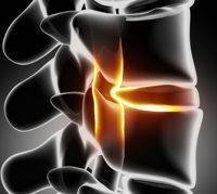
It may cause paralysis, and the patient must ensure the speed of accurate diagnosis, speed of treatment and follow-up of the herniated disc in the thoracic vertebrae ... Physical therapy helps relieve symptoms and strengthen muscles! undefined Some cartilage is exposed to erosion and may slip from its place Dr. Yasser bin Muhammad Al-Beheiri The middle part of the spine consists of twelve vertebrae called the thoracic spine, extending from the lower part of the neck and ending in the middle of the back over the lumbar region. These thoracic vertebrae are stacked on top of each other, and between them are the intervertebral discs. And that the thoracic vertebrae are connected and linked to the ribs, their movement is much less than that of the cervical and lumbar vertebrae, and therefore they are usually less prone to osteoarthritis and herniated discs. However, in spite of this, it may happen from time to time that one of these cartilages is exposed to erosion and may slip out of its place to give what is known as a herniated disc of the thoracic vertebrae. The importance of this type and this place of herniated disc is that the spinal cord passes through the thoracic vertebrae and is exposed to pressure on a small or large part of its parts as a result of the herniated disc. In addition, the slippage may cause blockage of the small arteries that supply the spinal cord, affecting its function.
Despite most cases of herniated disc in the thoracic vertebrae, it is the result of abrasion and roughness of the cartilage and affects the lower part of these vertebrae, but it may be the result of severe injury to the vertebrae and cartilage in traffic accidents, falls, or as a result of a sharp twisting of the mid-back region.
Symptoms vary greatly according to the severity of the herniated disc and the extent of the spinal cord’s impact, but usually it begins in the form of back pain centered in the area of the slipped cartilage and then extends to the sides in the mid-back region, and patients liken it to a rope or belt of pain surrounding them. In addition, the patient may feel tingling, burning, or numbness. As the disease progresses, symptoms may extend into paralysis, weakness and numbness in the legs, or even disturbances in controlling the outflow of urine and stool. In very severe cases, the herniated disc may lead to a large defect in the spinal cord that appears in the form of partial or complete hemiplegia. The diagnosis is usually made after a clinical examination in which the focus is on the neurological functions of the lower extremities, such as muscle strength, sensory effects, and the extent to which reflexes are affected and the functions of the vents. After that, specialized rays such as magnetic resonance imaging or CT scans are done, which usually show the location of the herniated disc, its size, the intensity of pressure on the spinal cord, and the presence of any changes in the spinal cord as a result of this pressure.
treatment
In cases where the symptoms are mild and there is no pressure on the spinal cord, conservative and non-surgical methods are usually used by prescribing medical belts, pain medications, anti-inflammatory drugs, taking several days to rest, avoiding fatigue and lifting weights. Additionally, physiotherapy sessions may help relieve symptoms and strengthen muscles. Also, oral cortisone medication may be prescribed for a short time if there are mild symptoms in the lower extremities. Usually the majority of herniated discs of the thoracic vertebrae respond to conservative treatment within a period not exceeding six weeks.
Surgical treatment
It is the best solution in cases that do not respond to non-surgical treatment or in cases where symptoms increase during conservative treatment or in cases where there is pressure on the spinal cord and affects the functions of muscles, exits or sensations. It is possible to remove the herniated disc in the thoracic vertebrae, either by microsurgery from the back or through an incision in the chest between the ribs. Usually, this surgery is preferred from the front through an opening in the chest because this provides a clearer view of removing the slipped cartilage and provides great safety to the spinal cord during surgery. In the past few years, new techniques have been developed to remove a herniated disc through video and a thoracoscopy or what is known as (Video Assisted Thoracoscopy Surgery). This helps in facilitating surgery, speeding recovery and rehabilitation.
Recommendations
Care must be taken to quickly and accurately diagnose in cases of herniated disc in the thoracic vertebrae, speed of treatment, accuracy of follow-up, speed of surgical intervention when there are symptoms indicating the presence of pressure on the spinal cord or the beginnings of paralysis in the lower limbs.