for Laparoscopic Cholecystectomy
In 1985, Prof Dr Erich Mühe of Germany performed the first laparoscopic cholecystectomy (LC) [1]. He performed 94 such procedures before another surgeon, Phillipe Mouret of Lyon, France, performed his first laparoscopic cholecystectomy in 1987 [2], who said: “Laparoscopy is the only method capable of performing a complete and valid surgical exploration of the abdomen, with the peritoneal cavity in nearly physiological conditions, except for the elevation of the anterior abdominal wall.” In 1988, the authors reported using this technique on 36 patients [2]. A gynecologist, William Saye, collaborated with general surgeon Barry McKernan to perform the first laparoscopic cholecystectomy in the United States [3].
Reddick and Olsen described their initial series of 25 patients in whom laser laparoscopic techniques were used to remove the gallbladder [4]. Several studies published in series, results supported decreased morbidity and reduction in hospitalization when compared to the traditional method for cholecystectomy. In 1990, my group performed the first laparoscopic under general anesthesia lasting more than four hours.
State-of-the-art 1990''s imaging technology was harnessed to visualize a diseased organ. Special instruments were conceived to manipulate these organs. Laparoscopic sutures and loops were refined along with clip appliers, staples, tackers and gastrointestinal staplers. Perhaps the most important of these developments was the laparoscopic clip applier, for this humble instrument gave inexperienced general surgeons the confidence that ducts and blood vessels could be quickly secured. Once this confidence was established, all else followed. The introduction of laparoscopic cholecystectomy proved to be a new era in the management of cholelithiasis.
Selection and Preparation of Patients
When laparoscopic cholecystectomy was first performed, only patients who were good surgical risks, with non acute disease and no prior abdominal surgeries were ed for the procedure [2]. However, as experience was gained, the pool of patients expanded to encompass those who were otherwise candidates for conventional cholecystectomy, and some surgeons included patients who had acute disease [5]. Current absolute contraindications are peritonitis, which could be aggravated by the laparoscopic technique [6]. Laparoscopic surgery was successfully performed in patient''s anticoagulated [7], during pregnancy [8] and morbidly obese [9]. Choledocholithiasis also has been considered a relative contraindication to the procedure because of the difficulty of laparoscopically removing stones in the common bile duct. However, newly developed instruments are useful for exploration of and stone retrieval from the common bile duct.
Laparoscopic Technique
Until recently the choice of anesthetic technique for upper abdominal laparoscopic surgery is mostly limited to general anesthesia with muscle paralysis, tracheal intubation and intermittent positive pressure ventilation (IPPV). At induction of anesthesia it is important to avoid stomach inflation during ventilation as this increases the risk of gastric injury during trocar ion. Tracheal intubation and IPPV ensure airway protection and control of pulmonary ventilation to maintain normocarbia. Ventilation with a large tidal volume of 12-15 mL/kg prevents progressive alveolar atelectasis and hypoxemia and allows for more effective alveolar ventilation and carbon dioxide elimination [10].
To perform the surgery laparoscopically, there is a need to a space between the abdominal wall and the viscera to allow adequate surgical team display and manipulation of abdominal contents. Although there are mechanical methods of abdominal distention, abdominal insufflation gas is a standard practice. The patient''s abdominal cavity is insufflated to a pressure of 10 to 14 mm Hg with 2-4 L of CO2 through a 1cm peri-umbilical incision. A laparoscope connected to a video monitor is then introduced through this incision to assist in accurate placement of three additional 5-mm to 10-mm subcostal cannulas evenly spaced from the midline to the right anterior axillary line. With the help of an assistant, subsequent manipulations are performed entirely through these four ports, with videoscopic guidance provided by the laparoscope. Blunt dissection is used to isolate the cystic duct and cystic artery. The cystic artery is double clipped and divided. Many surgeons then routinely perform cholangiography by clipping the cystic duct proximal to its junction with the common bile duct and ing a small catheter into the cystic duct through a small distal incision. When the anatomy is clearly defined after cholangiography, the cystic duct is then double clipped and ligated, and the gallbladder is dissected free from the liver bed by using either electrocautery or a laser for cutting and hemostasis. Once the gallbladder is dissected free from the liver, the laparoscope is usually switched to another port, and the gallbladder is pulled through the umbilical site, neck first, with a forceps. The CO2 is subsequently expelled through the ports by opening the cannulas, and the skin incisions are then closed with subcuticular absorbable sutures and sterile adhesive tape.
Monitoring
The electrocardiogram, noninvasive arterial pressure monitor, airway pressure monitor, pulse oximeter, end-tidal carbon dioxide concentration monitor, peripheral nerve stimulation and body temperature probe are routinely used. A urinary catheter is usually placed to minimize the risk of bladder injury and improve surgical exposure. The urine output should be monitored in patients with compromised cardiopulmonary function. The expired CO2 is easily monitored with an adapter placed in the nose. Persistent refractory hypercapnia or acidosis may require deflation of the pneumoperitoneum.
Regional Anesthesia
Until the last century regional techniques had not been reported for laparoscopic or surgical procedures in the upper abdominal region. Epidural anaesthesia has been used for outpatient gynaecological laparoscopic procedures to reduce complications and shorten recovery time after anesthesia. Local or regional anesthetic techniques have not been reported for laparoscopic cholecystectomy or other upper abdominal surgical procedures except in patients with cystic fibrosis. A high epidural block (T2-T4 levels) is required to abolish the discomfort of surgical stimulation of the upper gastrointestinal structures. The high block produces myocardial depression and reduction in venous return, aggravating the hemodynamic effects of tension pneumoperitoneum.
Many researchers have observed that performing laparoscopic surgery under regional analgesia carries many advantages. The reduction of surgical stress response is considered one of its major advantages. This is accomplished through two aspects the laparoscopic technique, itself which reduces the degree of tissue trauma and consequently the injury response (minimal invasive surgery concept), and the spinal analgesia itself which provides pain relief by blocking afferent neural block, together with block of various humoral mediator cascade systems [11]. Avoidance of airway instrumentation and lower incidence of deep vein thrombosis are other important advantages of this technique.
Surprisingly, in the era of minimally invasive medicine, regional anesthesia has not gained popularity in LC. Regional block such as low thoracic epidural [12], spinal [13], and combined spinal-epidural [14] blocks have been used in patients with relevant medical problems. This is due mainly to the notion that laparoscopic cholecystectomy requires tracheal intubation to prevent aspiration and respiratory complication due to the introduction of CO2 in the peritoneum [15], which would not be well tolerated by an awake patient during the procedure [16].
Spinal anesthesia has the advantage of providing analgesia and muscle relaxation with complete preservation of consciousness and rapid postoperative recovery. In addition, there is a protection against the potential complications of general anesthesia.
Spinal Anesthesia Technique
In general, laparoscopic procedures of the abdominal cavity necessitate endotracheal intubation and mechanical ventilation due to the induction of pneumoperitoneum. The increased intra-abdominal pressure together with the increased carbon dioxide load to the lungs are considered as better managed under mechanical ventilation, making thus general anesthesia a necessary requirement for these operations. In the past decade, a small number of reports appeared involving regional anesthesia for laparoscopic general surgery, including patients with coexisting pulmonary disease who were deemed high risk for general anesthesia. More recently, a limited number of studies showed the feasibility of the application of regional anesthesia on healthy subjects.
Reports for laparoscopic general surgery under regional anesthesia alone included patients with coexisting pulmonary disease, who are deemed high risk for GA. Recently, some studies compared to general anesthesia with spinal anesthesia for laparoscopic [17,18], with some advantages for regional technique.
After monitoring and venipuncture patients were monitored with non-invasive blood pressure, oxygen saturation, and expired CO2. An 18F catheter was ed in the left hand for hydration and administration of drugs.Fentanyl (1μg/kg) and midazolam (1 mg) were administered before the subarachnoid puncture.
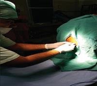
With the patient in left lateral decubitus or sitting position, after establishing aseptic conditions, the subarachnoid space was punctured between the L3-L4 apophyses with a 27G cut-bevel or pencil-point needle. Backflow of CSF confirmed the position of the needle in the subarachnoid space after the administration of 20 μg of fentanyl, 3 mL of hyperbaric bupivacaine were injected. Afterwards, patients were placed in the supine position with a 10-20 degree head-down. The stylet of the needle was used to test the lack of sensitivity of the patient, which should reach the level of T3. Once the goal was achieved, the surgical table was paced in the horizontal position and the patient was cleared for surgery.
Recently the anatomy of the thoracic spinal canal was investigated with magnetic resonance imaging in 50 patients [19]. It was also demonstrated the safety of spinal anesthesia with puncture at T10 using the combined spinal-epidural [20,21] or single puncture technique [22]. In one recent study of 300 patients, it was demonstrated that thoracic puncture using a cut point needle or pencil point needle was associated with the same incidence of paresthesia as was the lumbar approach, and without sequelae [23].
It is well known that anesthetists often fail to correctly identify the vertebral level. In 2000, the authors [24] investigated anesthetist''s ability to identify a marked lumbar space they showed that vertebral levels were identified correctly only 29% of the time. The T10 level was the landmark correctly identified by the largest number (92%) of anesthesiologists [24]. However, only 2% performed procedure thoracic spinal anesthesia [24].
Laparoscopic cholecystectomy has rapidly become a popular native to open cholecystectomy, and is considered a cost-effective technique for the treatment of symptomatic cholelithiasis. Spinal anesthesia has some advantages compared with general anesthesia [18,26], including the patient being awake and oriented at the end of the procedure, less postoperative pain, and the ability to ambulate earlier than patients receiving general anesthesia. The laparoscopic cholecystectomy may be performed at the level of thoracic puncture T10 [27]. The initial replacement and medications used are the same when performing lumbar puncture. In the case of thoracic puncture, it can be performed either in the later