

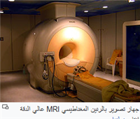
There are many different types today with many ideas for MRI devices. In general, there are three main types of MRI devices:
permanent.
resistant.
And anti-resistance.
The magnetic resonance device in general contains a part that gives a strong magnetic field and a part that emits radio waves to stimulate the protons and pick up signals coming from them, and a part of the gradient system.
A survey that is used in the medical field costs $1 million per Tesla and several hundred thousand dollars are spent annually on maintenance.
Computers are mainly used in MRI examinations, and their advanced programs effectively help give the best results.
Magnetic Resonance Physics
File:MR inversion recovery.ogvPlay media
The device consists of a huge spiral electromagnet to form a magnetic field around the patient, which produces a magnetic field of 2 Tesla, which is equivalent to 20,000 Gauss.
This field causes the hydrogen atoms to magnetize and all head to their north magnetic part, uniting in one direction. After that, the body is exposed to radio rays that lead to an increase in the energy of these atoms, and therefore it will change its direction to a certain degree, leaving us an atom out of every million atoms with which the magnetic resonance imaging process is done, which is a large number of atoms enough for the emergence of a clear picture of the part to be photographed and sends an amount of energy in reverse. This reverse energy is received from the device, calculated and formed in the form of an image. This image shows the intensity of hydrogen in each area of the body, and through this image doctors can discover many diseases.
When the atoms in the body are excited, the protons move with and against the direction of the main magnetic field. The protons corresponding to the main direction increase from the anti-protons by a small amount, but it is very important in obtaining the image later. It soon returns to equilibrium, but to return to equilibrium there are two important timings:
horizontal timing
Horizontal timing, which is the fastest timing, when protons are scattered on the horizontal axis and is symbolized by the symbol T2 on the horizontal axis, so it is called horizontal timing, which is a symbol of time, i.e. time, as well as vertical timing, the opposite of that
T2 relaxation.jpg
vertical time
The vertical time is the time when the protons return to their equilibrium position and is symbolized by the symbol T1
T1 relaxation.jpg
It should be noted that the two timings coincide with each other.
The energy of the magnet used in magnetic resonance is measured in tesla and is equal to 10,000 gauss. By measuring the average Earth’s magnetism, it was found to be equal to half a gauss.
Proton density
The number of active protons per unit volume of a tissue, and the density varies from one tissue to another.
echo cycle
After the impact of the protons with radio waves, the waves are transmitted again and return 180°, and the time period between the first effect is 90° and the second effect is 180° by echo time.
Spinecho.png
MRI uses
Beating Heart axial.gif
Mra1.jpg
The use of magnetic resonance imaging is for a diagnostic purpose such as imaging the veins and arteries, or imaging neurological changes in the brain, and magnetic resonance is considered the best type of imaging in clarifying tissues and body fluids, as well as used to plan treatment plans based on radiotherapy. Before the MRI examination, the medical history must be reviewed and it is absolutely sure that there are no previous surgeries or accidents that led to the presence of metals in the body such as shrapnel, and this is confirmed through a routine general x-ray examination & the patient’s passing through a metal detector. The patient is usually given a special dye that is injected into the body in order to increase the contrast and clarify the adjacent parts.
Magnetic resonance imaging
The magnetic resonance image consists of several columns and rows called matrix, each column and row contains squares called pixels, the signals received from the body are distributed on these squares so that they are arranged according to their arrangement in the body, and this mechanism depends on a gradient device that gives each slice of the body a force A specific signal, and the strength of the captured signal gives a color on the gray scale, so the MRI image is a grayscale image. The equation for special clarity is:
The number of squares per cm = 1/the volume of the square.
The contrast in the image depends on the horizontal and vertical timings and the density of the proton, which are called (internal effects), while the echo time and replay time are considered (external effects).
Clarity (IMAGE RESOLUTION)
Most of the magnetic resonance images consist of two dimensions, each dimension is divided into a grid consisting of rectangular image elements called (pixels).
The intensity of the image in each pixel depends on the strength of the magnetic resonance wave that is emitted from the region containing it.
Image size depends on the number of pixels, and most images consist of 265 pixels vertically and 256 pixels horizontally.
Definition of image resolution: It is the system''s ability to differentiate between two separate points.
The sizes of the voxels control the image resolution, as images with small voxels are well clear, thus making it easier for the system to differentiate between small body components.
Factors affecting image clarity and voxel size
slice thickness.
Display field FOV.
The number of the sham matrix.
Factors that control the field of view and voxel dimensions
The strength of the gradual magnetic field.
The sampling time (bandwidth) of the progressive field.
The number of the sham array can be increased by taking more samples (FREQUENCY ENCODING) and more phases of the progressive magnetic field in the direction (PHASE ENCODING).


