
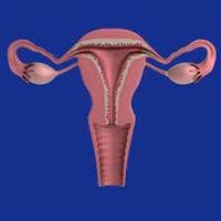
Cervical insufficiency is one of the most common causes of miscarriage, especially in the second trimester of pregnancy, as well as for premature birth. Cervical insufficiency means insufficiency of the internal cervical os.
Uterine insufficiency may be a congenital (birth) defect, but this form of uterine insufficiency is very rare, and it can affect women who were exposed, while in their mothers'' womb, to one of the types of drugs that were used thirty years ago, called Diethylstilbesterol - DES ).
Insufficiency is often the result of dilation of the cervix during a previous pregnancy, for the purpose of an intended abortion. During abortions, there is a need to dilate the cervix in order to empty the uterus of its contents.
There are two ways to dilate the cervix: The wrong image they make by means of metal expansion tools that are ed into the uterus, sometimes by force, which can cause damage to the elastic (soft) fibers in the cervix, causing shortcomings in the future.
The best method, used today in most hospitals that perform abortions, is to a small branch of Laminaria into the cervix a few hours before the process of dividing the uterus.
This algae has several hygroscopic properties, that is, it is able to attract water molecules, and expand gradually and slowly, so that it does not cause any damage to the endometrial orifice. In some very rare cases, cervical insufficiency occurs after a natural miscarriage, or damage to the cervix after a difficult delivery with the Vacum device.
Cervical insufficiency treatment
When cervical insufficiency is diagnosed, it is treated with cervical atresia (Cercloge). This procedure requires a simple and ingenious surgery first performed by an Indian gynecological surgeon named Shirodkar, and this process has been developed and improved over the years. In this method, an artificial suture made of a material called Marcelon (silk thread or any other material can also be used) is ed into the neck tissue, so that it encircles the neck approximately at the height of the ventral orifice, thus preventing cervical dilatation. When cervical insufficiency is diagnosed before pregnancy, this atresia is performed ively in the first weeks of pregnancy (12-15). But if the deficiency was not diagnosed before pregnancy, and this was discovered late through an ultrasound examination during pregnancy, it is possible to perform a late cervical atresia operation, but the chances of success of this operation are very few. Most likely, the stitch (suture) that was ed into the cervix is removed during the 38th week of pregnancy, after which the woman is often able to give birth naturally.