If your doctor is concerned that a brain tumor is causing your symptoms, a variety of tests may be conducted to aid in the diagnosis of a brain tumor and find out more about its size, location, and type.
Neurological exams and brain scans are used to detect the presence, location, and size of a tumor. Biopsies, molecular tests, and certain scans help determine the tumor type and provide other information, which may help determine treatment options.
As you go through your brain tumor journey, your doctor may repeat some of these scans and tests to find out if the tumor has changed and if it is responding to treatment.
Understanding these tests – how they work, why they are done, and what they show – can help you be an informed member of your care team. If you have any questions about the tests your doctor is ordering, ask your doctor, nurse, patient navigator, or other health care professional.
Neurological Exams
A neurological exam is usually the first step toward figuring out what is causing your symptoms. The goal is to see how well your nervous system is working. Because your brain does so many different things, a neurological exam can have many different components. Your doctor may repeat the neurological exam several times throughout your brain tumor journey to see whether your symptoms and functioning are changing.
During a basic neurological exam, your doctor evaluates how well your nerves, balance, reflexes, strength, and senses are working. Your doctor will also assess your cognitive functioning and mental or emotional state.
Here are some common parts of a neurological test
Mental Status: tests the awareness of person, place, time, and situation
Motor Function: tests the muscle power in various muscle groups to assess for any weakness
Sensory Exam: checks the ability to feel and if there is a difference between sides
Reflexes: uses a reflex hammer to gently tap different points on the body for movement
Cranial Nerves: the brain has 12 cranial nerves, each with its own purpose. These are checked to see if a specific nerve has been affected. There may be tests for smell, vision, facial sensation, hearing, swallowing, taste, shrugging shoulders, and tongue movement.
Balance and Coordination: checks if you are able to stand with your eyes closed, walk in a straight line, or touch your nose with your eyes closed
If neurological exam results lead your doctor to suspect that a brain tumor is causing symptoms, your doctor may order additional tests, including imaging or blood tests. Your doctor may also refer you to a neurologist (a physician who specializes in brain and central nervous system problems) or a neuro-oncologist (a doctor who specializes in cancer of the brain and the rest of the nervous system).
Brain Scans (Neuro-Imaging Studies)
A brain scan is a type of diagnostic test that takes pictures of the brain and/or spinal cord. Like an X-ray can see inside the body to diagnose a broken bone, a brain scan can see a tumor inside the skull. The two most common scans for diagnosing a brain tumor are magnetic resonance imaging (MRI) and computed tomography (known as a CT or CAT scan). A CT is often used to detect the presence of a tumor, and an MRI is used to gather more detailed information about the tumor’s size, location, and possible type (a biopsy is needed to confirm tumor type).
Brain scans are non-invasive. A noninvasive procedure is one that does not involve cutting into the body or removing tissue, and is generally painless. For some, the most challenging part of a scan is having to sit or lie still for a long time.
During a scan, your doctor may use a special dye, called contrast, to make areas of the brain easier to see. Contrast material is usually injected into a vein before the scan starts. Prior to the scan, you will be provided with information, including any details and instructions about the contrast agent (if prescribed), what to wear, and what to bring along. The doctor will determine if you need a blood test to ensure your kidneys are safe to receive contrast. Let the doctor know if you have any allergies.
Computed Tomography (CT)
CT scans are commonly used as screening scans, and are preferred for looking at bones or the presence of blood or abnormal brain tissue They are safe overall but do expose patients to a small amount of radiation. This scan combines high-speed X-ray technology with a computer. The CT scanner is a large donut-shaped piece of equipment. The patient lies on a table that slides into an opening in the CT machine. The CT scanner circles the head, taking X-rays from different angles. A computer then combines these X-ray images into a cross-sectional image of the brain (sometimes called a “slice”). A contrast dye may be injected to help the tumor appear more clearly in the CT scan results. These images help the doctor determine the presence and location of a tumor.
Magnetic Resonance Imaging (MRI)*
The MRI scanner is a tunnel-shaped piece of equipment that uses magnets and radio waves to generate two-dimensional or three-dimensional pictures of the brain tissue. These images are more detailed than those of a CT for assessing the presence of a brain tumor. The patient lies on a table that slides into the scanner, where a magnetic field surrounds the head. Prior to the scan, a contrast dye (typically gadolinium) is injected into the body. This contrast agent helps some tumors appear more clearly. The final images help the doctor determine the presence, size, and exact location of a tumor.
Functional MRI (fMRI)*
fMRIs produce MRI images that help identify parts of the brain responsible for important functions like speech or movement. This information can help a surgeon plan to remove a tumor without harming these functional areas. During this scan, you may be asked to answer questions or move certain parts of your body. These actions help identify the functional areas during the scan.
Flow-Sensitive MRI (FS MRI)*
This technique combines functional MRI with images of the flow of cerebrospinal fluid (CSF), a colorless liquid that fills and surrounds the brain and the spinal cord. FS MRIs are particularly useful in planning surgery to remove a skull base tumor, spinal cord tumor, or a tumor causing hydrocephalus (fluid buildup on the brain).
Perfusion MRI *
The type of scan works by seeing how blood moves through the brain. It can help to determine if a brain mass is a tumor or something else (e.g., abscess, stroke, radiation related change from prior radiation, or cells killed by radiation called radiation necrosis). It also helps predict what type of tumor it may be, or whether new growth of a tumor is actually tumor or if it is radiation necrosis or pseudoprogression.
CT Angiography (CTA) and Magnetic Resonance Angiography (MRA)*
These scans are used to locate blood vessels in the brain. This information can help the surgeon plan for tumor removal. This scan may be done with a contrast dye.
Magnetic Resonance Spectroscopy (MRS)*
This type of MRI provides information about the chemical make-up of brain tissue. It helps determine tumor type (which must be confirmed by biopsy), identify dead tissue (also called necrosis), or the difference between old and new tumor cells.
Positron Emission Tomography (PET, FDG-PET)
A PET scan shows how your brain is working as opposed to just the structure of the brain. Before the test, a small amount of radioactive material, (called a “tracer”), is injected into the bloodstream. The tracer will then collect into areas with actively dividing cells. Tumor cells are actively dividing so they will show up as bright spots on the scan. It may also help your doctor estimate the tumor’s grade and in some cases, it may help to tell the difference between recurrent tumor cells and necrosis (cells killed by radiation). The tracer leaves the body quickly and does not pose a safety risk to you or your loved one.
Single Photon Emission Computerized Tomography (SPECT)
SPECT is not usually used to diagnose brain tumors. However, it sometimes provides data that complements information captured in other scans. This data includes how malignant the tumor may be and the difference between tumor tissue and cells killed by radiation (necrosis).
For these tests, the radiology facility will ask you to complete a form determining if they are safe. For instance, metal objects implanted in your body or certain medical conditions like kidney disease can pose safety risks.
Tumor Biopsy
A biopsy is a surgical procedure in which the surgeon removes a small amount of tumor tissue. This tissue sample is commonly collected during brain surgery to remove a tumor. A biopsy may be performed on its own when surgery is either not necessary or not possible, but more information about the tumor is needed.
The purpose of a biopsy is to establish an exact tumor diagnosis. After it is collected, tumor tissue is sent to a lab for a pathologist (a physician who interprets and diagnoses diseases) to evaluate. The pathologist views the tumor tissue under a microscope to determine the tumor type and grade. Specialized stains and molecular testing provide additional information to help with diagnosis.
There are two types of biopsies, open and stereotactic
Open Biopsy
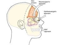
During a resection (surgical removal of a tumor), the surgeon takes the tumor tissue sample while the tumor is exposed. This generally requires the surgeon to remove a piece of the skull to access the tumor.
Stereotactic Biopsy
While under general anesthesia, the surgeon drills a small hole (called a burr hole) into the skull using a computer-assisted guidance system. This system uses 3D images and data from a CT or MRI scan to provide precise information about a tumor’s location and its position relative to the brain’s many structures. A thin, hollow needle is ed through the hole and collects a sample of tumor tissue, called a “core”, through the needle.
Pathology Reports
A pathology report is a document that summarizes key information about your tumor. A pathologist (a physician who interprets and diagnoses the changes caused by disease in tissues and body fluids) examines and analyzes the tumor tissue after a biopsy. These results not only provide a diagnosis of your tumor, but also help doctors determine the status of your tumor and plan treatments.
Here are common parts of a pathology report
General information:patient’s name, date of birth, medical record number (MRN), doctor’s name, specimen case number, date collected
Location: g. brain, frontal lobe
Gross description: tumor size and what the tumor looks like to the naked eye
Microscopic description: what the Pathologist sees under the microscope and how it compares with normal cells
Diagnosis: provides the final pathology of the tumor and includes the tumor type, grade, molecular markers, and possibly the mitotic rate (how quickly the tumor cells are dividing).