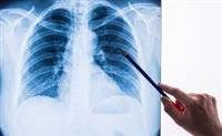
A chest radiograph or chest radiograph is an imaging test that visualizes the rib cage by releasing x-rays. The rays pass through the chest and are absorbed by a plate that contains a sensitive tape for this radiation. The examination is performed by an x-ray technician, and the image is transferred to the doctor on film or in a computerized form, and the doctor in turn analyzes it.
It is worth mentioning that the doctor who specializes in analyzing radiological images is a radiologist, and doctors specializing in other fields also analyze radiological images, and consult a radiologist when needed.
The rib cage normally contains the following
Bones such as the ribs and clavicle.
the lungs.
Respiratory ducts.
the heart.
major blood vessels.
Therefore, diseases that affect these organs can sometimes be diagnosed by radiography, and other organs that are not in the chest area can also be photographed, but chest imaging is not intended for diagnosing diseases in these organs, such as:
The upper part of the stomach.
diaphragm.
the neck.
shoulders.
One of the main advantages of chest imaging is that it is available, has limited exposure to radiation, and is cheap. Its main drawback is its limited sensitivity compared to other imaging methods.
Therefore, chest radiography is used almost routinely in many medical conditions, and its results can help the doctor choose additional examinations.
When is the examination performed?
The examination helps to find the cause of many cases, the most prominent of which include the following:
Finding the cause of common complaints such as coughing, shortness of breath, or chest pain.
Diagnosis or evaluation of certain lung diseases such as pneumonia, asthma, tuberculosis, and lung cancer.
Diagnosing or evaluating fractures or diseases of the bones of the rib cage, such as a rib fracture.
Diagnosis of injuries in the event of an accident.
Determine the location of foreign bodies that absorb radiation, such as rings that were swallowed or pulled out and stuck in the esophagus or in the trachea.
After an invasive procedure in which tubes or catheters were ed in order to determine the location of the tube that was ed, and if there was a need to correct its location.
category at risk
There is little risk to fetuses of pregnant women and children, as they are more sensitive to radiation.
Despite this, the degree of radiation resulting from this examination is very limited compared to all other radiological examinations. However, when concerned, the doctor who recommended imaging should be consulted to discuss the need for it.
Diseases related to chest imaging
The most prominent diseases related to this examination include the following
lung diseases.
heart disease.
Fractures of the ribs and clavicle.
Perforation in one of the abdominal organs (perforation).
Pneumothorax.
Hemothorax.
Lung cancer and lymphoma.
The method of conducting the examination
Traditional chest imaging is usually done from the back and from the side, but there is the possibility of imaging while sitting or lying down. the chest.
The imaging technician installs the plate in its designated place, directs the x-ray tube from the other side, and asks you not to move, as movement affects the quality of the image. It may require taking a deep breath.
In the event of pregnancy, you must inform the doctor who recommended the examination, as well as the radiology technician. If your medical condition requires a chest x-ray, the pregnant woman is provided with a lead gown to cover the abdomen.
Analysis of the results
Proper chest X-ray analysis involves
No infiltration, effusion, or congestion.
Heart reflection intact.
No bone fractures.
Central trachea.
If there is a breathing tube in case the patient is breathing artificially or a nasogastric tube, it must be in place.
Improper chest X-ray analysis includes
Leakage or effusion: It may indicate a lot of diseases, but most often the talk revolves around infection.
Enlarged lymph glands: which can result from many diseases, including tumors.
Pulmonary congestion: often indicates pulmonary edema.
The shadow of an enlarged heart: It often indicates a heart problem, but it may result from a technical problem when imaging.
Flat diaphragm: indicates a problem in the small airways such as asthma or chronic obstructive pulmonary disease (COPD).