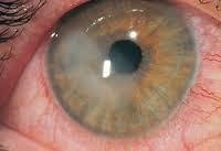

Keratoconus treatment depends on the degree of disease progression, from installing special contact lenses to corneal transplantation. Keratoconus is an eye disease that affects the cornea, also called (Kera conus). In conjunction with progressive convexity, the cornea also becomes thin. The conical and asymmetrical shape of the cornea causes asymmetric refraction
The light rays are inside the cornea, so the light rays are not focused well on the retina. Then, the resulting image on the retina becomes blurry and blurry.
This process usually begins in the late teens or twenties, and can develop slowly over several years. Keratoconus can affect the cornea in both eyes or in one eye. Apparently, keratoconus is a genetic eye disease. So far, few genes have been found that cause this disease.
Symptoms of keratoconus!
Keratoconus can develop slowly over several years, but it may develop rapidly, too. The first signs of this include a rapid rise in the number of glasses, especially the rise in the cylinder, which sometimes develops and becomes greater than the number of glasses itself. This increase is the result of an increase in corneal curvature and the increasing asymmetry in its structure, as things appear blurred and distorted. In addition, increased sensitivity to light and dazzle can develop.
Sometimes keratoconus is suspected during an eyeglass examination at the eyeglasses manufacturer, where the patient is directed to an examination by an ophthalmologist.
What are the reasons for the development of keratoconus?
There are many causes of keratoconus, most of which are hereditary and some are acquired.
The genetic causes of keratoconus are still unknown, for the most part. So far, there have been few genetic defects that cause keratoconus, but there may still be many defects to be discovered. In some cases, keratoconus appears in a number of family members, especially in families where consanguineous marriage occurs.
Research indicates that there is a weakness in the corneal tissue as a result of an increase in the activity of enzymes that break down the protein that builds the cornea. This process exposes the cornea to oxidative damage by free radicals. As a result, the corneal tissue becomes thinner and thinner, and the pressure inside the eye causes the cornea to protrude outwards, distort it, and asymmetrical curvature.
Acquired factors for keratoconus include excessive rubbing of the eyes, wrong choice of contact lenses, allergic eye infections and chronic inflammatory conditions of the eye.
How is keratoconus diagnosed and treated?
Keratoconus is diagnosed through a combination of tests.
Slit lamp examination - a device based on a microscope that allows examination of the eye at high magnification. In cases of advanced keratoconus, corneal convexity and thinning can be distinguished. In cases of advanced keratoconus, fine lines parallel to each other can be seen in the inner part of the cornea, and scarring is sometimes seen in the center of the cornea.
Corneal topography - This examination is the first method of diagnosing keratoconus. This examination consists of imaging the cornea and building a topographical map of the corneal surface. The map shows with relative accuracy the curvature of the cornea at each point. The color table shows the different bumps. In a normal cornea, the convexities are uniform, so the terrain color is uniform. In keratoconus, one area can be seen in which there are too many bumps, as this area is outlined in an orange-red color.
Corneal topography is a quick, easy to perform, and non-invasive examination. The examined person puts his head against the device, and within about a minute a colored picture of the cornea is obtained. With this examination, the progression of corneal curvature can be accurately tracked.
Corneal thickness test - This test is based on corneal ultrasound. This examination, too, is short and non-invasive, and gives a measurement of the thickness of the cornea at different points of it. Everyone has a different corneal thickness, as the average human corneal thickness is 530 microns. In people with keratoconus, the thickness of the cornea is expected to be less than 500 microns, and in advanced cases it can also be less than 400 microns.
Keratoconus treatment
Treatment of keratoconus includes several stages, and depends on several factors such as its severity, the degree of development of keratoconus upon diagnosis, and the patient''s vision ability.
When it comes to a mild degree of disease, glasses or soft contact lenses can provide a solution to improve vision. But as the disease progresses and the cornea becomes more asymmetric and curved, reasonable visual acuity cannot be obtained with glasses or soft contact lenses.
How are corneal transplants performed?
Treatment of moderate and advanced keratoconus includes the following methods
• Implantation of rings in the cornea – rings consisting of two semicircular sections, which are implanted in the cornea, on its periphery (Fig. 12). These rings are made of a solid material, and by installing them in the cornea, they make changes in the shape of the cornea and reduce its curvature.
Keratoconus treatment with orthogonal nodes - a new treatment method based on the formation of orthogonal nodes between collagen fibers in the cornea. Collagen is the protein that builds up the cornea. The underlying assumption behind this treatment is that by strengthening and tightening the corneal tissue it will be possible to stop the process of its curvature, especially if the treatment is carried out at the stage of appearance of keratoconus, or the beginnings of its development.
• Treatment of keratoconus with corneal transplantation - Corneal transplantation is suggested as a last resort in keratoconus, when it is not possible to wear contact lenses, or when contact lenses cannot provide reasonable vision. The transplanted cornea is obtained from a donor. Due to the small number of organ donors, there is a problem with the availability of corneas for transplantation, and as a result, the waiting time for corneal transplantation is usually not short. But, nevertheless, corneal transplantation for keratoconus, in general, is an operation with great chances and possibilities of success. The cornea does not contain blood vessels, and therefore it is not exposed to the immune system, like other organs. Therefore, the risk of corneal rejection after successful transplantation is low compared to other transplants.