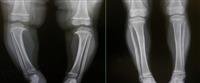
The treatment of fractures in children differs from that in adults, as children have a number of distinct phenomena that in turn affect the treatment of fractures, such as: a great ability to grow and build bones, and the ability to spontaneous repair.
Injury to the growth plates at the ends of the bones can cause serious growth disturbances. Therefore, the growth plates must be monitored and monitored by X-ray images when any fracture occurs near them.
Symptoms of common fractures in children
Symptoms can occur slightly differently in each child. Here are the most common symptoms if your child breaks a bone:
pain.
swelling.
Obvious bone deformity.
Trouble using or moving the bone in a normal way.
Warmth of the fracture site.
flushing
bruises
numbness.
Obvious bone deformity.
These symptoms may sound like other health problems So make sure your child sees his healthcare provider for a diagnosis.
Causes and risk factors for common fractures in children
The following is an explanation of the most prominent causes and risk factors for common fractures in children:
1. Common causes of fractures in children
Children love to run, jump, skip, and stumble, all of which are activities that can lead to a forearm fracture in the event of an unexpected fall. In most cases, forearm fractures in children occur because of:
Fall on an outstretched arm.
Direct fall on the forearm.
A direct blow to the forearm.
2. Risk factors for common fractures in children
The most important factors include the following
Genetic factors.
Malnutrition including inadequate dietary calcium intake, avoidance of milk, and excessive consumption of soft drinks.
Lack of weight-bearing physical activity.
Obesity.
Higher exposure to trauma may affect the risk of fractures in general.
Common fracture complications in children
The most prominent complications include the following
Damage to blood vessels
Many fractures cause significant bleeding around the injury. Rarely is bleeding inside the body internal bleeding, or from an open wound external bleeding large enough to cause a potentially life-threatening in blood pressure.
Nerve damage
Sometimes nerves are stretched, bruised, or crushed when a bone is broken. A direct blow can bruise or crush a nerve. These injuries usually heal on their own over weeks to months to years depending on the severity of the injury. Some nerve injuries do not heal. completely.
pulmonary embolism
Pulmonary embolism is the most common acute complication of serious hip or pelvic fractures. It occurs when a blood clot forms in a vein, breaks off, becomes an embolus, travels to the lung, and blocks an artery there. As a result, the body may not get enough oxygen.
Fat embolism
Fat embolism rarely occurs as it can occur when long bones, such as: the femur, are fractured and fat is released from within the bone. Fat may travel through veins and settle in the lungs and block blood vessels there, causing pulmonary embolism.
compartment syndrome
Compartment syndrome rarely occurs. For example, it may develop when affected muscles swell too much after a broken arm or leg. Because swelling compresses nearby blood vessels and blood flow to the affected limb is reduced or blocked. As a result, the limb''s tissues may be damaged or die and the limb may have to be amputated.
infections
If the skin is torn when a bone is broken, the wound can become infected and the infection may spread to the bone.
joint problems
Fractures that extend into a joint usually damage the cartilage at the ends of the bones in the joint.
Osteonecrosis
When blood flow to the bone is disrupted, part of it may die, leading to osteonecrosis.
Diagnosis of common fractures in children
The most prominent methods of diagnosis include the following
1. Physical examination
After discussing your child''s symptoms and medical history, your doctor will perform a careful examination of the fracture site to determine the extent of the injury. He or she will look for:
A deformity around the elbow, forearm, or wrist.
The patchy place.
swelling.
Inability to rotate or move the fracture site.
During the physical examination, your doctor will make sure that the nerves and blood circulation in your child''s hands and fingers are not affected.
2. Radiography
Include the following
X ray
X-rays provide clear pictures of dense structures such as bone, and since the hand, wrist, arm, and elbow can all be injured during a fall onto an outstretched arm Your doctor may order x-rays of the elbow, wrist, and forearm to determine the extent of the injury.
MRI
This test uses a combination of large magnets, radio frequency, and a computer to make detailed pictures of the organs and structures inside the body.
CT scan
This test uses X-rays and a computer to make detailed pictures of the body. A CT scan shows details of bones, muscles, fat, and organs. This type of imaging is more detailed than a general X-ray.
Treating common fractures in children
Therapeutic methods include the following
Treatment for forearm fractures depends on the type of fracture and the degree of displacement. Your doctor will use one or both of the following treatments to treat a forearm fracture:
1. Non-surgical treatment
Some stable fractures may simply need the support of a cast or splint while they heal.
For more severe fractures that have become tilted, a doctor may be able to manipulate the bone or gently push it back into place without surgery. This procedure is called a closed reduction, after which the arm is immobilized in a cast or splint while it heals.
2. Surgical treatment
In some cases, surgery is needed to align the pieces of bone and hold them in place. Your doctor may recommend surgery if:
The bone has penetrated the skin and this type of injury is called an open fracture and is at risk of infection and requires specific treatment.
Bones cannot be properly aligned through manipulation alone.
The bones have already begun to fuse at an angle or in an improper position.
Your doctor will open the skin and reposition the broken bone fragments. Your doctor may use staples, metal implants, or a cast to hold the broken bones in place until they heal.
Prevention of common fractures in children
Although some children are more susceptible than others, prevention methods can be followed, which include the following:
Your child gets enough calcium.
Exercising to strengthen bones. Weight-bearing exercises, such as jumping rope, jogging, and walking, can help develop and maintain strong bones.
Common types
The most common types of fractures include
Clavicle fracture
The collarbone, the tapering, curved bone that extends from the top of the middle breastbone to the shoulder blade, may be damaged in a child at birth, or during infancy when a child falls on an outstretched arm.
Head of humerus fracture
The injury occurs when falling while the arm is extended backwards and the elbow is straight. In the examination, any nerve injury should first be ruled out.
Supracondylar fracture
These fractures account for 60% of supracondylar fractures in children, with 97% of these fractures occurring in a fall while the elbow is straight, usually at the age of 5-8 years.
These fractures are more common among males, and the fracture site is above the articular surface of the arm bone at the elbow, and the ends of the bones may injure the blood vessels and nerves that pass near them, and therefore the pulse and nerves must be examined before treatment.
Lateral condyle fracture
It accounts for 17% of elbow fractures, especially in children at the age of 6 years, as this injury occurs when falling while the arm is outstretched and the elbow is compressed inward.
Fractures of the medial epicondyl
These fractures constitute about 10% of elbow fractures in children at the age of 9-12 years, as this injury occurs when the arm is stretched and the elbow is pressed outward as the epicondyle separates from its place, and this separation may also occur when the elbow is dislocated. out of place.
Head of radius fracture
It constitutes about 10% of elbow fractures where the head of the radius may move from its place. The type of treatment is related to the extent of the displacement. If the displacement is up to 30 degrees, the treatment is conservative, while if the displacement is greater than that, then there is a need for reconstruction under general anesthesia and then splinting.
Forearm fractures
It constitutes about 45% of all fractures in children over the age of 5 years, as this injury occurs when falling while the arm is stretched together with a twist, as x-ray imaging is done in order to detect additional injuries.
Greenstick fracture
The compressed part of the bone usually suffers partial fractures, while the taut part remains complete and intact. Treatment is usually done by splinting with a splint placed for three weeks.
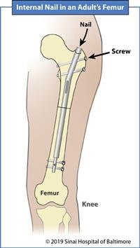
Fractures of the femoral shaft
These fractures occur as a result of a direct injury, such as an accident or a fall, or as part of child abuse.
Tibia/fibula fractures
Common among children of all ages, about 50% of these fractures occur in the distal third of the leg, and are treated by splinting with gypsum for a period of six weeks.
Ankle fractures
These fractures pass through the growth plates in the shin bone and the fibula bone, so the reconstruction must be accurate, and usually an open reconstruction is required in addition to internal splinting, and the growth plates should be closely and continuously monitored for fear of injury or deformation in the aftermath.