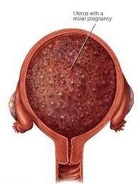
A molar pregnancy is a non-cancerous tumor that arises inside the uterus. A molar pregnancy occurs when the placenta develops into a group of follicles and does not develop into a healthy pregnancy with healthy placental tissue and a fetus.
In a complete molar pregnancy, there are no healthy placental tissues at all, and there is no fetus either. On the other hand, in a partial molar pregnancy, there are few healthy placental tissues in addition to unhealthy fetal tissues, and therefore a live fetus cannot be formed.
A molar pregnancy may turn into a rare type of cancerous tumor, and therefore this condition must be addressed urgently, as careful follow-up of the course of molar pregnancy is important in order to reduce the complications of this problem and to allow an opportunity for a healthy pregnancy to occur in the future.
Symptoms of molar pregnancy
Pregnancy appears in the early stages of molar pregnancy as a healthy pregnancy, but after that symptoms of molar pregnancy begin to appear, which are:
Vaginal bleeding that appears in the first trimester of pregnancy and is often dark brown to bright red in color.
severe nausea and vomiting
The exit of a cyst that looks like a grape.
A feeling of pressure or pain in the pelvic area.
Very rapid enlargement of the uterus, meaning that the size of the uterus is very large in relation to the size of the uterus that is supposed to be at this stage of pregnancy.
high blood pressure.
Preeclampsia.
Ovarian cysts.
Anemia.
Hyperthyroidism.
A thick, cyst-filled placenta that fills the uterine cavity.
Causes and risk factors for molar pregnancy
In a healthy pregnancy, the egg is fertilized from the mother by the sperm from the father, and it contains 46 chromosomes, 23 chromosomes from the mother''s egg and the remaining 23 from the father''s sperm.
1. Causes of molar pregnancy
Causes of molar pregnancy include
The source of all chromosomes is from the father in a complete molar pregnancy due to the loss of chromosomes from the mother and duplication of the paternal source chromosomes.
In a partial molar pregnancy, there is no loss of chromosomes that come from the mother, but a double set of chromosomes that come from the father, meaning that the fertilized egg contains 69 chromosomes.
A partial molar pregnancy occurs when there is a duplication of the number of chromosomes that come from the father, or after the fertilization of one egg by two different sperms.
2. Risk factors
There are several risk factors that lead to a molar pregnancy
Age: Hydatid pregnancy is more common in pregnancies that occur in women age 35 and over or girls younger than 20.
Previous estrous pregnancy: The probability of a estrous pregnancy occurring in women who have previously had a estrous pregnancy is about 10 times higher than that of other women.
Race: Hydatid pregnancy is more common in women of Southeast Asian descent.
Complications of molar pregnancy
In 10% of cases, after aborting a molar pregnancy and expelling the diseased placental tissue, hydatid tissue remains in the uterus, which continues to grow. This can be seen when a blood test is performed, as it shows high levels of the pregnancy hormone, and vaginal bleeding occurs if the hydatid tissue penetrates the layers of the uterus.
Hydatid tissue develops very rarely into choriocarcinoma and may spread to other organs in the body. Choriocarcinoma treatment also depends on the combination of chemotherapy and is usually successful.
Diagnosis of molar pregnancy
In an ultrasound examination that is preferable to be performed at this stage via the vagina, it is often possible to note the absence of a fetus, the absence of amniotic fluid, and other symptoms.
The following must also be done
Check blood strength.
Checking the level of thyroid hormone.
Preeclampsia examination.
Treatment of molar pregnancy
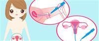
There is no possibility that a molar pregnancy will turn into a healthy pregnancy, and therefore the molar pregnancy must be removed as soon as possible, as the pregnancy is removed by a surgical operation that can be performed under local anesthesia or general anesthesia, as the duration of the surgery is short and most often lasts for 15 minutes up to 30 minutes.
The operation is done through the vagina and does not require an abdominal incision. In the event that the volume of hydatid tissue is large and the woman has no desire to have children, the uterus can be completely removed. After the operation, the level of pregnancy hormone in the blood is monitored. To ensure that the hydatid tissue has been completely removed.
After the hormone level returns to the normal level, follow-up of the case continues for half a year and up to a year. During this period, the doctor may advise not to become pregnant to ensure that the increase in the pregnancy hormone is not caused by the development of the hydatid tissue again.
This condition may require chemotherapy and is generally successful.
Prevention of molar pregnancy
There is no way to prevent, but a woman who has previously suffered from a molar pregnancy must be monitored regularly by a doctor to avoid infection later.