Have you ever heard of cholangioscopy? Learn with us in this article the most important information about this topic.
In this article, we will talk about cholangiopancreatography, or what is known as endoscopic retrograde cholangiopancreatography, and the most important things related to this topic:
What is cholangioscopy?
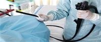
It is a surgical procedure used to diagnose and treat obstruction in the biliary system. In this procedure, an endoscope equipped with a camera is placed in the mouth and ed until it reaches the duodenum to image the bile ducts, gallbladder, pancreas, or liver.
This procedure is usually used to obtain a biopsy, repair, or remove blockages in these bile or pancreatic ducts.
When is a cholangioscope used?
Bile duct obstruction is one of the most important reasons that require cholangioscopy. Stones are the most common cause of bile duct obstruction. Other reasons for using cholangioscopy are the following:
Bile duct stricture.
Inflammation of the bile duct.
Acute pancreatitis.
Chronic pancreatitis.
The presence of surgical complications in the bile ducts or pancreas.
Pancreatic pseudocyst.
Bile duct tumors.
Pancreatic tumors.
Some liver diseases.
Symptoms that may make the doctor suspect the presence of diseases in the bile ducts are the following:
stomach ache.
Itching.
Jaundice.
Vomiting and nausea.
Pale or off-colored stools.
Dark urine.
What tests may a patient undergo before bile duct endoscopy?
The patient may undergo some of the following tests
Abdominal ultrasound.
Computed tomography examination of the abdomen.
Percutaneous transhepatic cholangiogram.
Magnetic resonance cholangiopancreatography.
How is bile duct and pancreas endoscopy performed?
Bile duct endoscopy is performed in the following steps
The patient must fast the night before the endoscopy procedure.
The patient must be given general anesthesia and given painkillers.
Antibiotics may be given to patients who take immunosuppressants or in whom complete relief of bile duct obstruction is not expected.
The patient should lie on his left side.
The doctor numbs the end of the throat with a local anesthetic by spraying it.
The endoscope is passed into the stomach and then into the duodenum, where the opening of the common bile duct is located.
The doctor may pump air into the duodenum to improve the quality of the imaging.
The doctor injects contrast material using a wire passed through the endoscope through the opening of the common bile duct.
An X-ray image is taken, and these images indicate areas of blockage.
The endoscope may be used for more invasive procedures, such as removing a stone, ing a stent, or taking biopsies.
The patient will begin to wake up hours after the endoscopy, and then the doctor will monitor the patient''s vital signs and ask about pain or discomfort.
The patient may need painkillers during this period. If the patient''s condition is stable, he may be able to return home a few hours after the procedure.
What are the possible complications of cholangoscopy?
Some patients may suffer from complications of cholangioscopy, but the rate of these complications is small, and these complications include the following:
An allergic reaction to the intravenous dye used during x-ray imaging.
An infection of the gallbladder or bile duct.
Perforation of the stomach, bile ducts, or small intestine.
pancreatitis.
Internal bleeding occurs.
Warning signs that may indicate the occurrence of complications include the following
body''s temperature raising.
Severe abdominal pain.
Jaundice.
Frequent vomiting.
Cough accompanied by blood.
Vomiting blood.
Blood in stool.