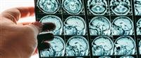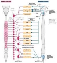
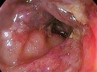
Neuroscience is a modern branch of radiology, which specializes in the treatment and diagnosis of diseases affecting the central, peripheral, nervous, and nervous system.
This science is divided into two parts Interventional Neuroradiology and Diagnostic Neuroradiology.
Interventional Neuroradiology is called another Endovascular Neurosurgery, and it is a relatively new medical specialization and is concerned with treating brain diseases, head, neck, and spinal cord diseases with techniques that lead to minimal harm or pain, and thus produce It means a complete recovery from diseases that could not be cured 15 years ago.
As for the diagnostic neurological radiology, it is also one of the modern sub-specializations of radiology, and it represents the methods used in the diagnosis of diseases of the nervous system, head and neck to enable the specialist of radiological interventional radiology to treat such diseases. It has witnessed a great development so that specialists can now reach the accurate diagnosis and make vascular treatment interventions without even having to make an incision in the skin at times.
Interventional Neuroradiology
This specialization started in the eighties of the twentieth century at the hands of a neurologist and neurosurgeon, and it took rapid development simultaneously with the development in medical technology in general, where the latter is one of the important factors that accelerated the development of this relatively modern science.
Over time, the techniques and devices developed by the interventional neurologist in the last five years have led to the treatment of more brain diseases that threaten their lives with high efficiency. This modern science represents an important transitional stage in the role of radiologists from consultants only to actual doctors who play an active role in the therapeutic process and with a large level of responsibility.
This early specialization helped radiologists and surgeons to come up with new techniques to treat patients who could not be treated with traditional methods such as open brain surgery, for example. For medical conditions and diseases that have hindered traditional treatment, they include giant cerebral aneurysm, inoperable aneurysm, large arterial venous anomalies, and other difficult health problems that coincide with neurological diseases. As an native to the traditional treatment of such patients, the introduction of Cerebral Angiography gave way to their treatment more effectively.
Among the diseases that can be cured using interventional neurological techniques to achieve a faster recovery of patients, reduce the length of stay in the hospital, and expose them to a lesser degree of risk posed by surgeries
Aneurysm, by ing platinum coils into the dilated vessels to stimulate their clotting and avoid their bursting.
Strokes.
Enlarged arteries in the brain by injecting a substance that prevents bleeding.
Vascular facial tumors.
Fractures of vertebrae.
Diagnostic neurological radiation
Diagnostic therapeutic rays include the use of medical devices and devices and radiography to diagnose diseases of the nervous system, head and neck, and thus enable doctors to treat them.
This subspecialty of diagnostic radiology began in 1927 with the release of the first vascular imaging report in which specialists were able to see the anatomy of the brain vessels in a person alive, after making a small surgical incision in the neck to puncture the carotid artery, and then injected a shadow contrast material to the rays with the aim of Sequential cinematography of the arteries and veins of the brain.
Tens of years later, vascular imaging began to evolve significantly to become safer, more effective and accurate, as the surgical incision was initially canceled and the perforation of the carotid artery directly through the skin, and then the process followed became the cutaneous epithelial catheter, by ing the catheter tube during The femoral artery after percutaneous puncture, as well as the use of safer contrast agents.
In the nineties, important developments were made in the angiography process. The first was Digital Subtraction Technology, which reduced the need to use traditional methods of developing films, which consumed a lot of time after each time the contrast material was injected.
There is also Roadmap Fluoroscopic Imaging technology that enables the catheter tube to be directed to the correct place within the blood vessel, thereby disseminating the material necessary to treat aneurysm in the brain or inflating a balloon in these vessels to plug them.
In addition to the previous two techniques, many factors later contributed to making angiography a safe and effective method when used by experts and specialists, such as the development of mechanical devices used to inject contrast materials and the development of high-performance catheters tubes.
Simultaneously with the developments in vascular imaging, there have also been developments in computerized tomography and magnetic resonance imaging, and now computed tomography angiography-cta and magnetic resonance angiography-mra (imaging) Cerebrovascular.