
Mammography examination is one of the most important methods of early detection of breast cancer. The most important information about it is in the following. Through the following:
It contributes to the early detection of diseases in the breast, by using a special X-ray imaging machine. This examination is not invasive, but it leads to external pressure on the breast.
Mammography examination allows for the possibility of carrying out diagnostic examinations, screening examinations, as well as monitoring the treatment of tumors.
It is used as a method for biopsy and labeling before surgery.
Mechanism for conducting a mammogram examination
During a routine radiographic examination, two images are taken of each breast from two different angles, that is, four images for each patient in total. As for the mammogram examination that contains implants (which are external materials implanted for the purpose of breast augmentation), it includes eight images, four images for each breast.
It is recommended to perform the examination using digital devices, as they allow a better view of the various details, and the radiation levels in these devices are low, especially when it comes to digital devices.
The images obtained through the examination are sent to the specialist doctor to analyze the results, and the images are kept for a long time so that it is possible to compare the conditions of the breast in different periods of time, and the comparison includes the changes that occur in the breast tissue, even if it is very slight.
Early diagnosis of the disease provides the possibility of recovery in more than 85% of cases, and the possibility of diagnosing the disease at an early stage depends on the ability of the specialist doctor to analyze the results of the mammogram examination.
Terminology related to mammogram examination
In the following, we provide an explanation of a group of terms used during a mammogram examination:
breast
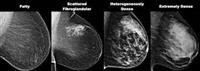
It consists of adipose tissue (dark in color) and glandular tissue (light in color). The denser and richer the glandular tissue, the more difficult the mammography examination is to perform, and the greater the need for a complementary ultrasound examination.
However, there are many things that can only be discovered through a mammogram examination, such as: some cases of calcification.
Pectoralis muscle
It is the muscle on which the breast rests.
Axillary lymph nodes
Part of the body''s defense system against infections and foreign cells, including cancer cells.
Cystic fluid mass
It is always a benign mass.
Solid mass
It is a mass that can be benign or malignant. The mammography examination cannot distinguish between the types of masses, so it is followed to perform complementary examinations, such as: ultrasound examination, fine needle aspiration examination (FNA-Fine Needle Aspiration), and needle biopsy examination Thick (CNB-Core NeedleBiopsy).
Calcification
It is a phenomenon that occurs as a result of the deposition of calcium in small amounts in the breast tissue. Calcification can be seen in the pictures in the form of small points.
But usually it is not possible to feel it, and this phenomenon is known especially after menopause. In fact, this does not necessarily have to be cancer, but it could be one of the indicators that indicate the presence of cancerous activity.
There are two types of calcification: macrocalcification, which is the deposition of calcium in large quantities in the breast tissue, and microcalcification, which is the deposition of calcium in small amounts. These deposits can appear in the form of groups or shapes that indicate accelerated cellular activity.
In these cases, complementary examinations are performed, such as: biopsy examination, ultrasound every few months, and sometimes calcifications have nothing to do with the cancerous process but with old injuries, dilated milk ducts, calcium deposition in the fluid of a benign cyst or swelling as a result of injury.
However, 49% of carcinoid tumors detected by mammogram appear as calcifications.
Scleroderma
It is a phenomenon related to breast engorgement and this phenomenon occurs in the aftermath of mastitis, cancer, lymphatic drainage duct damage, heart failure, ovarian enlargement or skin condition after radiation therapy to the breast.
Skin thickness can be measured with a mammogram or ultrasound.