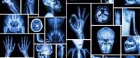
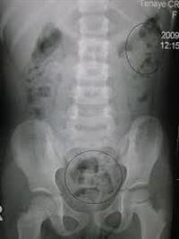
Abdominal radiography is a group of medical imaging tests that gives us an image of the abdomen through X-ray imaging called the Roentgen. The rays pass through the abdomen and penetrate it to be picked up by a board that contains a special imaging tape that is sensitive to this type of radiation. This examination is performed by an x-ray technician, and the image is transmitted to the doctor
The specialist takes the form of an image or sends it as a computerized image for analysis. The doctor responsible for analyzing the radiology results is usually a radiologist, and a radiologist is consulted when needed.
The special and important advantages of abdominal imaging is that it is almost always available, the amount of radiation to which the patient is exposed is small, and it does not cost a lot of money.
When is the examination performed?
Due to the special characteristics of the internal organs in the abdominal region, abdominal imaging may be suitable for diagnosing only a limited number of emergency cases, and not many pathological conditions.
Therefore, this examination is common and widespread mainly in emergency rooms in order to ensure that there are no conditions that can be diagnosed. An abdominal imaging examination is also performed in the following cases:
Determine the cause of abdominal pain, nausea, and constipation.
Find the location of the bowel obstruction.
Finding the location of the perforation in an organ of the gastrointestinal tract.
Finding stones in the stool, which is usually caused by chronic constipation.
Finding stones in the kidneys or in the urinary tract.
Finding foreign objects that absorb radiation, such as: metal pieces that have been swallowed or ed through the anus.
It can be done after invasive examinations, which include the introduction of a tube or catheters, in order to estimate the location of the tube that was ed, and if there is a need to adjust its location.
category at risk
It is likely that there will be little risk in the process of imaging the abdomen in fetuses in pregnant women who undergo this examination, as well as young children, especially since this group is usually more sensitive to radiation.
Radiation levels are higher during imaging of the abdominal region than during imaging of the chest area, for example, but they remain lower than radiation levels in some other examinations, such as: computed tomography examination, therefore, for these pregnant patients and children, it is recommended to perform an ultrasound examination to photograph the abdominal region.
related diseases
Diseases related to this examination include the following
Intestinal Obstruction.
Perforation in one of the organs of the digestive system (Perforation).
Tumors of the gastrointestinal tract.
Ulcers in the stomach and duodenum (Ulcers).
chronic constipation
Kidney stones.
Inflammatory bowel diseases.
The method of conducting the examination
Regular abdominal imaging is performed in two modes
Abdominal imaging while lying on the back.
Photographing the abdomen while standing.
For patients who cannot stand, they are asked to lie on their side.
Most of the time, the x-ray technician asks the patient to take off his clothes and remove jewelry that may hinder the imaging process, and then the technician puts the imaging board in the place designated for him, and the device that emits x-rays is directed from the other side, asking the patient not to move because movement It may damage the image quality.
Analysis of the results
Possible results of this examination include
1. Proper results of an abdominal imaging examination
Usually, if the results of the abdominal imaging examination are correct, they will not be detailed.
Usually, no growths or abnormal amounts of fluid (Ascites) or foreign bodies are seen. Normal amounts of air and fluid in the intestine appear on proper abdominal imaging, and the amount of stool is within reason in the large intestine.
2. Improper results of an abdominal imaging examination
There are many results that are not considered improper in an abdominal imaging examination, including:
A blocked bowel. This is because part of the bowel is larger than normal or areas in the bowel have abnormal amounts of air or fluid.
The gathering of air inside the abdominal cavity, but outside the intestines, and it is caused by a hole in the stomach or intestines.
The appearance of the walls of the intestine abnormally or thick.
The size, shape, or location of the bladder or kidneys may be abnormal, and kidney stones may be seen in the kidneys, ureters, bladder, or urethra.
Gallstones can be seen on an abdominal X-ray.
Some tumors can be seen.