
The development of magnetic resonance imaging (MRI) was the result of the continuation of human curiosity that led it to the technological progress that has brought us to today.
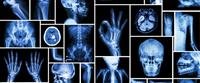
Human curiosity led to the development of X-ray imaging, also called Alentgen, which is the first method that made it possible to look inside the living human body, but there were several drawbacks to X-ray imaging, most notably:
Inability to see the soft tissues.
Radiation can cause cancer.
This prompted researchers to look for additional ways to look inside the human body.
MRI is one of the advanced methods that allow looking inside the living human body, and one of the advantages of this method is that it does not use radiation that can cause damage, as in X-ray imaging, so it can be used safely in examining pregnant women.
In the MRI examination, strong magnets and radio waves are used, so that the magnet arranges the hydrogen atoms in the body, which are the most common atoms in the human body, in one level, and then the radio waves are sent towards the body, which leads to a change in the direction of these atoms.
When you stop emitting radio waves, these atoms are arranged again in one plane, where the computer measures the time period it takes for these atoms to return to the same plane and then uses this information to form the image.
One of the significant advantages of this method is that it does not use ionizing radiation, which can cause mutations and cancerous tumors.
The main drawback of this method is the inability to use it to examine people who use a metal pacemaker or who have metal pieces in their bodies, due to the use of very strong magnets.
When is the examination performed?
As in X-ray imaging, this examination is also performed in order to examine one of the internal organs. The difference between these two examinations is in the type of organs that can be examined.
Whereas X-ray imaging forms an image of hard tissues in the body such as bones, MRI s an image of soft tissues as this examination is mainly used to image the brain, spine and joints, and is used, for example, to check whether there is a disc herniation in the spine.
However, due to the high cost of this test, it is recommended to use it only when no other method is available to diagnose the problem.
category at risk
An MRI scan is particularly successful for examining and imaging the following organs
the brain.
vertebral column.
joints.
This examination can be performed in people who suffer from problems in the mentioned organs.
In addition, this examination can be used to visualize the heart, liver, and other internal organs when needed. It is preferable for people who feel uncomfortable and uncomfortable in enclosed spaces to take sedatives before undergoing this examination, due to the type of examination that requires lying on a bed that is ed in A place like a cave, which is the magnet itself.
related diseases
There are many diseases and conditions that necessitate the use of an MRI scan, and among the most common cases are the following:
Suspected disc herniation in the spine.
Suspicion of a brain injury, such as a hemorrhage or a brain tumor.
Diseases of the nervous system, such as: multiple myeloma.
Joint injuries, such as meniscus rupture.
The method of conducting the examination
An MRI examination is a non-invasive and painless examination, and is performed by the following:
Remove jewelry during the examination.
The clothes are required to be taken off if they contain metals such as buttons or zippers, and the patient is given other special clothes.
It is required to lie on a bed that is ed into the device that contains magnets, the examination takes between 30-40 minutes.
The person being examined is asked not to move Because movement s a blurred image, during the examination the person may sometimes feel a little warm in the examination area and this is very normal.
During the examination, the person hears light knocks when the device takes pictures, and in most places where this examination is performed, earphones can be requested in case of disturbance from the noise.
It should be noted that it is possible to return to normal and routine activities immediately after the examination is completed.
Analysis of the results
After the results of the MRI examination are issued, the radiologist examines them, and he is a doctor who specializes in analyzing medical images. Several days pass until the results of the examination are obtained, due to the need for accurate analysis of the images.
On the other hand, when the examination is performed in the hospital following an emergency, the results are obtained within a short period of time.
The examination report contains the names of the various organs that were examined, and it is indicated next to each member whether signs of disease were found in it or not.
After receiving the results, you should go to the treating doctor to determine how to continue treatment, according to the results.