breast cancer
Amidst the increase and the high number of women with breast cancer, there is no hope for technological progress that helps in early diagnosis as well as in treatment.
Advanced technology and early detection of breast cancer
3D mammogram, 4D ultrasound, MRI-guided biopsy, and advanced technology devices developed in recent years help detect and treat breast cancer in its early stages, offering great and convenient solutions for patients!
The rate at which breast cancer is growing is accelerating each year and it is one of the most common types of cancer. Despite this frustrating aspect, there is some good news as well. Although the incidence of breast cancer has increased, the death rate associated with breast cancer has decreased, and early diagnosis of the disease is very helpful in better treatment.
The advanced technological devices used in the detection and treatment of breast cancer play a fundamental role in this good development. Dr. Gül Esen İçTen, radiologist at Acibadem Maslak Hospital, provides information on some of the modern technology devices used in the detection and treatment of breast cancer and their advantages:
3D mammography
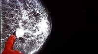
3D mammography is one of the latest and most important equipment provided by advanced technology. Actually, it is a digital radiography. The device itself and the way the image is taken is very similar to a regular mammogram.
How is it applied? During the radiation, the x-ray tube inside the machine moves from right to left, and a series of images are taken from different sections of the breast with a thickness of 1 millimeter by using a very low dose of radiation, the total radiation dose received in this process is equivalent to the dose received In a normal mammogram, the 3D mammogram must be performed with a normal mammogram.
What are the features of this photography?
These include
Cancer has become easier to spot: breast tissue made up of supportive fat tissue and mammary gland tissue can mask cancerous tissue. Especially if there is excess mammary gland tissue and supporting tissue (dense breasts), cancerous masses can hide behind this type of tissue. On the other hand, the exact opposite can happen when breast tissue crowds together to cause an appearance that looks like cancer, but is not cancer. A 3D mammogram is not limited to a single still image, but it can take a series of images of the breast. Therefore, the risk of misdiagnosis is greatly reduced.
Avoiding unnecessary procedures: Increasing correct diagnosis reduces the need for unnecessary imaging, biopsies, and operations.
Offers a more comfortable imaging position: The most uncomfortable aspect of a mammogram for a woman is the pressure on the breast in the machine and the pain and tightness that occurs because of this, while the breast is not pressed with the same force in a 3D mammogram, it is less painful for the woman.
Errors caused by non-expert people are reduced to a minimum.
3D ultrasound
Also called automatic ultrasound, volumetric ultrasound and 3D ultrasound, in this method a series of pictures are taken of the breast. This device is especially useful for medical centers that do not have the possibility of having a doctor check the results at all times.
How does it work?
Operated by a Technician, Not a Doctor The latest technical development in breast ultrasound known as 3D ultrasound takes 15-20 minutes, where an image of the whole breast is obtained conventionally and the image sets are evaluated by the doctor on a special monitor. The images obtained can be reconfigured at any segmental plane and measurements can be taken, and the doctor can reassess questionable areas later.
What are the features of this examination?
Highlights include
Parts of the breast are not overlooked So the errors are reduced to a minimum.
Traditional mammograms are taken, so it is easier to make comparisons when following the patient.
It is characterized by accurate, goal-oriented diagnostics.
Biopsy under MRI guidance
Breast MRI examination is used in women diagnosed with breast cancer to determine the presence of any other foci or not before surgery and to search for cancer in women who have a high risk. Suspicious findings not visible with any other imaging modality that can be seen with MRI In many cases, an MRI-guided biopsy must be performed to diagnose suspicious findings, which are only visible on MRI.
How is it implemented?
During the MRI-guided biopsy, which is a procedure that can only be done in a limited number of centers today, the patient lies face down as with all mammography cases, and with the help of a computer, a three-dimensional identification of the suspicious area is made, and a needle biopsy is taken from this area or are scheduled for surgery.
What are the features of this examination?
Highlights include
Avoiding Unnecessary Operations: In the not too distant past, it was not possible to biopsy tumors that could only be seen by MRI in Turkey. These patients are either invited for frequent check-ups, while they wait anxiously, or they are subjected to unnecessary surgeries. This is the only way to make Biopsies are possible with all suspected tumors.
He managed preventive breast surgery: when there were unrecognizable lumps in the breast, it was not previously possible to carry out preventive treatments for women suffering from breast cancer, and it was usually necessary to remove the entire breast. This problem is now solved Because a breast MRI makes biopsies possible for all tumors.
The sample is taken in 30 seconds.
Vacuum biopsy
Vacuum aspiration biopsy is the most advanced of the needle biopsy methods and is generally used to screen for tiny calcifications that are only visible on a mammogram and can be the first indication of breast cancer. In addition to microcalcifications, vacuum biopsy is notable as the method of choice for biopsies of all suspicious visual findings on mammogram and MRI, as it is not possible to excise all of the suspicious segment using this method.
Of course, if the patient is diagnosed with cancer, she may still need regular surgery to clean the area more extensively. Because there may be small concentrations of residual cancer that cannot be seen on imaging, this method makes it possible to completely remove small polyps without the need for surgery.
How is a suction biopsy performed?
A suction biopsy is done on a special table made especially for this procedure. It has a hole in the middle through which the breast is suspended. There is an X-ray tube in this table, which takes pictures of the suspicious area similar to a mammogram from different angles.
These images help to determine the three dimensions of the suspicious area. The biopsy is performed by a radiologist from under the table. A suction device and a disposable biopsy needle are used for the suction biopsy. The needle is placed according to the dimensions calculated by the computer. During the procedure, the suspicious area is withdrawn. It is continuously inside the needle where many tissue samples are taken.
What are the features of this examination?
Scan features include
The patient does not see the procedure or feel any pain.
It is possible to extract more tissue samples in half a minute than with other methods. Therefore, the correct diagnosis rate is 97-99%.
It is possible to excise any small suspicious areas, and it is a method that can be considered especially as an native to operations for benign tumors.
Single-dose radiotherapy during surgery.
LIAC device
Radiation therapy is provided to breast cancer patients in a specially protected room located in the radiation oncology departments by special devices that produce photons and electrons called linear accelerators. Radiation therapy continues for 5-7 weeks, according to the patient''s condition.
Patients come for treatment every day except for Saturday and Sunday, as the device used is a device that produces only electrons and is located in the operating rooms, and radiation therapy is completed in one dose during the operation.
How is it implemented?
After surgical removal of the tumor from the breast, the special part of the device is placed on the tumor area, and after taking the necessary measurements, the high dose of radiation is focused on this area only with complete protection of healthy tissue.
Although this single-dose method has been widely applied all over the world in recent years, it is not suitable for every patient. The radiation oncologist, surgeon, radiologist, and pathologist, who are directly responsible for the patient''s care, must decide together. Whether this method is suitable for the patient.
What are the advantages of this treatment?
Highlights include
Radiotherapy is done entirely during surgery.
The high dose of radiation is given only to the affected area, healthy tissue is protected.
It is not necessary to go to the hospital every day after surgery, and there is time for the wound to heal and for planning the next steps, and this provides an important advantage for the patient.