

styloidectomy: How should Eagle syndrome be managed surgically?
A.S. Al Weteid, M. Miloro: Transoral endoscopic-assisted styloidectomy: How should
Eagle syndrome be managed surgically?. Int. J. Oral Maxillofac. Surg. 2015; 44
1181–1187. # 2015 International Association of Oral and Maxillofacial Surgeons.
Published by Elsevier Ltd. All rights reserved.
Case Report
Oral Surgery
A. S. Al Weteid1, M. Miloro2
1Riyadh Dental Center, King Saud Medical Complex, Riyadh, Saudi Arabia; 2Oral and Maxillofacial Surgery, University of Illinois, Chicago, IL, USA
Abstract. Eagle syndrome, or calcification of the stylohyoid ligament, is a rare condition that may present a clinical diagnostic dilemma for those unfamiliar with its existence and its typical presenting signs and symptoms. Management of this disease process may involve either non-surgical or surgical treatment options. When surgery is indicated, the choice of a specific surgical modality is highly variable and is generally dependent upon individual surgeon preference and experience, since the location of the styloid process is consistent between patients, and the required surgical access is also similar depending upon the specific surgical plan. This paper reports a case of Eagle syndrome managed with a transoral endoscopic-assisted approach, explores the advantages and disadvantages of each surgical approach, and reviews the literature regarding surgical management options for Eagle syndrome.
Key words: Eagle syndrome; styloid process;
calcified stylohyoid ligament.
Accepted for publication 31 March 2015
Available online 16 May 2015
The styloid process is a thin bony protu- berance that emerges from the skull base. It has several attachments including the stylomastoid foramen, the jugular fora- men, the mastoid process, and important- ly, the carotid canal.1 Typical styloid process lengths range between 1.0 and
3.0 cm, and styloid processes of more than
3.0 cm are defined as excessively long, although in the articulated, or pseudo-ar- ticulated cases, each segment of ossified stylohyoid ligament may be less than
1.0 cm. Bilateral styloid process elonga-
tion is the most common presentation, but
interestingly, clinical symptoms are usu- ally present only unilaterally.2 The stylo- hyoid ligament is a fibrous band that attaches to the most caudal aspect of the styloid process and the cranial aspect of the hyoid bone (lesser cornu). Mineraliza- tion of the stylohyoid ligament occurs infrequently and is usually only an inci- dental finding noted on routine radio-
graphic examination. Eagle syndrome is diagnosed only when the facial or neck pain originates from a stylohyoid ligament that is calcified. This clinical presentation was identified in 1937 by W.W. Eagle.1
Eagle syndrome is divided into two sub- types: classic and carotid. The classic type exists when a calcified stylohyoid liga- ment results in facial, neck, and throat pain, possible dysphagia, globus sensa- tion, and tinnitus. The pain may radiate to the ipsilateral ear, and turning the head to the opposite side while simultaneously swallowing can reproduce the painful symptoms. These symptoms certainly evoke a wide differential diagnosis and this may make the diagnosis of Eagle syndrome very challenging from a clinical perspective. Also, due to the proximity of
0901-5027/0901181 + 07 # 2015 International Association of Oral and Maxillofacial Surgeons. Published by Elsevier Ltd. All rights reserved.
the stylohyoid ligament to the internal carotid artery, a vascular form of Eagle syndrome (carotid type) may occur, with carotid artery compression on head turn- ing, peri-orbital pain, and symptoms of transient ischaemic attacks or stroke (diz- ziness, headache, or syncope).
The clinical patient examination may allow palpation of an elongated styloid process, or bimanual palpation transorally and transfacially may localize the pain to the retromandibular region. If the pain is reproduced with turning the head to the contralateral side and swallowing simul- taneously, there should be a heightened index of suspicion for the presence of Eagle syndrome. From a radiographic per- spective, Eagle syndrome may be a com- mon incidental finding on routine panoramic radiographs, although only a very small percentage of patients with calcified stylohyoid ligaments present with clinically significant symptoms. Cur- rently, the use of computed tomography (CT) or cone beam CT (CBCT) scanning, with or without three-dimensional recon- struction, is essential to establish an Eagle syndrome diagnosis and to appropriately plan individual patient management.
Once the diagnosis of Eagle syndrome
has been established, there seems to be little consensus regarding the appropriate treatment algorithm to follow.3–14 Most clinicians will begin with non-surgical options for management using pharmaco- logical therapy with a variety of medica- tions, including non-steroidal anti- inflammatory agents, glucocorticoids, anticonvulsants, and local anaesthetic injections for symptomatic relief of the
painful symptoms. Once the decision has been made to treat the patient surgi- cally, there is controversy regarding the most appropriate approach to consider. In general, the position of the calcified and elongated stylomandibular ligament is consistent between patients, therefore the anatomical relationships do not vary a great deal and an individualized ap- proach is not necessary based upon ana- tomical location alone.
Since there is no consensus in the lit- erature regarding the most appropriate surgical approach to manage Eagle syn- drome, the purpose of this study was to review the literature in order to determine whether there is a preference for a trans- oral or extraoral approach to the elongated stylohyoid ligament in Eagle syndrome, and whether this preference is based upon the specific surgical specialty group in- volved. In addition, a novel approach to this controversial topic is presented: the case of a patient with Eagle syndrome
treated using an endoscopic-assisted trans- oral approach is reported; this maximizes the advantages (e.g. the use of a familiar surgical approach) and minimizes the dis- advantages (e.g. limited surgical access) of the transoral approach, and minimizes the potential untoward consequences of an extraoral transcervical approach (e.g. unaesthetic facial scar and facial nerve injury).
Materials and methods
A 33-year old Caucasian woman pre- sented to the Department of Oral and Maxillofacial Surgery of the University of Illinois complaining of pain in her left submandibular region that had started 5 years previously, with a history of left- sided neck pain on head movement and swallowing. The patient had consulted several head and neck specialists, includ- ing dentists, otolaryngologists, and oral and maxillofacial surgeons, in an attempt to determine the cause of her symptoms as well as to resolve her complaints. Al- though none of the specialists could deter- mine the aetiology of her pain despite the presence of classic signs and symptoms, as well as radiographic evidence on prior panoramic radiographs and CT scans of the presence of calcified stylohyoid liga- ments, she was diagnosed with myofascial pain by one specialist and treated with non-steroidal anti-inflammatory drugs and physical therapy without effect. She then underwent a tonsillectomy and ade- noidectomy 2 years later by another spe- cialist in an attempt to alleviate her pain, without any effect. The patient described the pain as a chronic, vague, generalized pain and a sensation similar to a ‘‘. . .tooth- pick under my jaw bone with pain that radiates to my left ear.’’ In addition, she reported mild dysphagia, sore throat, and functional limitations in her neck move- ment with resultant pain. She denied any discomfort or symptoms from her right side. In addition, she did not have a history of bruxism, which could theoretically lead to calcification of the stylohyoid liga- ments. On physical examination, the pain- ful area was localized to the retromandibular region with bimanual pal- pation transorally and transfacially, and the calcified stylohyoid ligament was pal- pable deep in the left tonsillar fossa. The pain was exacerbated by turning the head to the contralateral (right) side and simul- taneously swallowing.
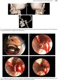
A CBCT scan demonstrated nearly
complete calcification, without pseudo-ar- ticulation, of the left stylohyoid ligament, almost to the level of the hyoid bone, as
well as partial calcification of the stylo- hyoid ligament on the right side (Fig. 1). Based upon the history and clinical examination, the decision was made to manage this case of Eagle syndrome sur- gically with a transoral approach, since prior non-surgical management with non- steroidal anti-inflammatory drugs had been ineffective and since the styloid pro- cess was palpable transorally. The patient was taken to the operating room for trans- oral resection of the elongated calcified stylohyoid ligament. An intraoral incision was made (left lateral posterior buccal vestibular approach, similar to a sagittal split osteotomy incision) and a sub-peri- osteal dissection was then carried out on the medial aspect of the mandible with blunt dissection (Fig. 2A). The medial pterygoid muscle was identified and blunt- ly dissected. Once this dissection was completed, the stylohyoid ligament was identified posteromedially (Fig. 2B). Careful blunt dissection was continued using a 4.0-mm zero-degree endoscope to expose the calcified stylohyoid liga- ment. Two vessel loops were passed around the ligament to retract the superior and inferior tissues and to isolate the ligament. After complete and clear iden- tification of the entire calcified ligament, the ligament was excised from its superior aspect, near the skull base, with a sharp rongeur (Fig. 2C). Dissection was then carried out inferiorly to detach all soft tissues from the caudal aspect of the liga- ment, and the calcified ligament was then removed in to (Fig. 2D). The length of the resected portion of the ligament was ap- proximately 4.0 cm, and the removal was documented with a postoperative CBCT
scan (Fig. 3).
Results
Postoperatively, the patient experienced an immediate relief of the symptoms she had had prior to surgery, including com- plete resolution of the left-sided subman- dibular pain as well as the left-sided neck pain during swallowing and turning of her head to the opposite side. Considering these clinical findings, this procedure of endoscopic-assisted transoral resection of the styloid process resulted in a clinical success. At the 6-month follow-up visit, the patient reported no recurrence of her pre-surgical symptoms.
Discussion
Eagle syndrome was documented by W.W. Eagle,15–19 and is defined as symp- tomatic mineralization and elongation
Fig. 1. Three-dimensional reconstruction and sagittal CBCT images showing the elongated calcified stylohyoid ligament: (A) left styloid process, (B) right styloid process, (C) sagittal of left styloid process.
Fig. 2. Intraoperative endoscopic views: (A) the incision, (B) identification of the calcified ligament, (C) resection of the cranial portion of the ligament, and (D) completed removal of the elongated calcified stylohyoid ligament.
Fig. 3. (A) Specimen of the calcified stylohyoid ligament. (B) Postoperative three-dimensional reconstruction showing the resected stylohyoid ligament with a small portion remaining at the skull base.
One common regimen is to use hydrocor- tisone 25 mg diluted with 1 ml 0.25% lidocaine injected in close proximity to the styloid process or painful area. Anti- epileptic preparations are also used, and
400 mg of carbamazepine per day is used initially, with increased dosage as tolerat- ed for effect for 2–3 weeks. As an - native to carbamazepine, and without the adverse side effects, gabapentin 300 mg per day, tianeptine 1.5 mg per day, and tramadol hydrochloride 37.5 mg/acet- aminophen 325 mg 3 tablets per day has been used.26 Also, a combination of ste- roid and local anaesthetic agent (triamcin-
olone–mepivacaine) can be injected in the tender regions. Subsequently, a stellate ganglion block may be performed once per week for approximately 4 weeks.26
Other non-surgical treatment methods in- clude pain reduction through physical therapy via relaxation of the associated muscles in the area that has been stretched, constricted, or in spasm as a result of stimulation of the stylohyoid ligament by the calcification process.25
The surgical treatment methods involve either fracture or excision of the elongated styloid process via an intraoral or extraoral approach. The two most common surgical approaches described in the literature in- clude either transoral manual fracture of the calcified stylohyoid ligament, or resec- tion of the stylohyoid ligament via a trans- oral or transfacial surgical approach. The rationale for inducing a fracture of the ligament and allowing retraction either in a cranial or caudal direction is that there is immediate relief of the pressure on the remaining areas of the ligament, which will act to relieve the painful symptoms. Regarding the two surgical approaches (transoral and transcervical), there are
(ossification, calcification, or pseudo-ar-
ticulation) of the stylohyoid ligament. In fact, Eagle syndrome was first described in
1652 by an Italian surgeon, Pietro March- etti, and the clinical signs and symptoms were defined by Eagle.1 The first surgical procedure done for the relief of symptoms associated with styloid process elongation was in 1872.3 From a demographic stand- point, patients with Eagle syndrome are generally in the fourth to sixth decades of
or long-acting local anaesthetic injections
may be used in the tonsillar region or individual clinically symptomatic area- s.20–25 One of the main components of treatment is the use of glucocorticosteroid hormones, which remain a mainstay of non-surgical therapy for these patients in order to decrease the inflammation of the ligament leading to the painful symptoms.
advantages and disadvantages of each sur-
gical technique (Table 1).
Although many incisions may be used for the transoral approach, a sagittal split osteotomy incision is useful since it is a well-known approach for most oral and maxillofacial surgeons, and it avoids a blind incision and dissection into an area with vital neural and vascular structures. A
life, although it can occur in adolescents and the elderly. Eagle syndrome is twice as common in women as men.
Both non-surgical and surgical treat-
ment methods exist for the management of Eagle syndrome. For non-surgical treat-
Table 1. Comparison of the intraoral and extraoral surgical approaches to the styloid process for
Eagle syndrome.
Intraoral approach Extraoral approach
No external scar Cutaneous scar
Shorter operative time Increased operative time
Restricted operative field Risk of facial nerve injury
ment, oral medication with gabapentin,
antidepressants (e.g. amitriptyline), and anticonvulsants (e.g. valproic acid, carba-
Increased risk of deep cervical infection (clean-contaminated wound)
Better visualization of the surgical field
Decreased risk of deep space neck infection
mazepine) can be used, and corticosteroid
(clean wound)
sub-periosteal dissection is carried to the medial pterygoid muscle, which is either retracted or divided to access the styloid process, and this dissection proceeds prox- imally and distally along the stylohyoid ligament, with surgical resection of the majority of the elongated calcified stylo- hyoid ligament, which helps to prevent recurrence.
The intraoral approach is favoured from an aesthetic standpoint since it avoids an external incision and resulting neck scar. In addition, the transoral approach may result in the potential for shorter operative times due to accelerated exposure and wound closure. One of the potential dis- advantages of the transoral route is expo- sure of the pharyngeal spaces to the intraoral clean-contaminated environment which elevates the risk of infection, and, therefore, perioperative antibiotics are used routinely. Further potential disadvan- tages of the transoral access include diffi- culty with carotid artery exposure and control in the event of iatrogenic intraop- erative injury, oropharyngeal and hypo- pharyngeal airway oedema, and
postoperative trismus. With the endoscop-
On the other hand, with the external approach, an incision is made in a skin crease midway between the mandibular angle and the mastoid bone. The sterno- cleidomastoid muscle is retracted posteri- orly, as well as the parotid gland superiorly, and the digastric muscle (posterior belly) inferiorly. This surgical approach may re- quire more time than a transoral approach, with dissection through the fascial layers of the neck and with possible intraoperative facial nerve monitoring, since this approach certainly places the facial nerve at risk of injury, and transient paresthesia may be expected from neurapraxic traction injury. The styloid process is identified and isolat- ed and freed from surrounding soft tissue attachments and distal connections to the hyoid bone. The styloid process is resected partially or completely, and the wound is closed in a layered fashion, with a resulting cutaneous neck scar. The advantages of this external approach include an improved op- erative field exposure and decreased risk of infection since it is considered a clean wound.
Table 2 shows the results of an evalua- tion of the literature from 2001 to 2014 regarding the surgical management of Ea- gle syndrome with a transoral or transcer- vical approach.27–47,23,48,5,49,50,22,51–54 Of
the 31 articles identified and 92 patients treated, there was a slight preference for the transoral approach (50 cases) over the transcervical approach (42 cases). Only one study reported a complication of the transcervical approach in two of five patients who experienced a transient weakness of the facial nerve (marginal mandibular branch).22 This review sup- ports the fact that the decision regarding
the use of a transoral or transcervical approach is surgeon-dependent and not necessarily based upon a clearly identifi- able risk–benefit ratio. This literature re- view does not assist in the decision- making process for the use of a transoral or transfacial approach, since there is a near even preference and there are very few reported complications. Additionally, we were not able to identify a specialty- based preference (Otorhinolaryngology
ic-assisted approach, if significant bleed- ing is encountered, initial treatment should involve pressure and the cautious use of the endoscope to identify a vessel at the point of severance, and either suture liga- tion or electrocoagulation, if possible. A
Table 2. Intraoral vs. extraoral approach for Eagle syndrome (literature 2001–2014).
Transoral/
Author [Ref.] Number transcervical Complications
1 Torres 2014 27 11 Transoral –
2 Bertossi 2014 28 1 Transcervical –
3 Dong 2014 29 1 Transcervical –
30
contingency plan should include wider surgical access either with extension of
the transoral incision, or an additional
4 Moon 2014
5 Bensoussan 2014 31
6 Ferreira 2014 32
1 Transcervical –
1 Transcervical –
1 Transcervical –
transcervical incision to gain vascular ac- cess. If transoral access alone is not suc- cessful, then the most appropriate management of a significant vascular bleed encountered during an endoscopic- assisted approach is to obtain pressure
hemostasis and utilize interventional radi-
7 Kim 2014 33 3 2 Transoral –
1 Transcervical –
8 Kar 2013 34 1 Transcervical –
9 Hoffmann 2013 35 1 Transcervical –
10 Thotappa 2012 36 1 Transoral –
11 Matsumoto 2012 37 1 Transoral –
12 Bahgat 2012 38 1 Transoral –
13 Todo 2012 39 2 Transcervical –
40
ology with arteriography to identify the bleeding site, with embolization of the vessel(s) involved. In addition, with the
14 Valerio 2012
15 Koivuma¨ki 2012 41
16 Santini 2012 42
17 Mayrink 2012 43
1 Transcervical –
1 Transoral –
1 Transoral –
1 Transcervical –
likelihood of mild to moderate airway
oedema from a transoral approach, bilat- eral procedures should not be performed simultaneously. Lastly, full exposure and excision of the entire calcified stylohyoid ligament may not be possible, but excision
18 Nakagawa 2011 44 1 Transcervical –
19 Yavuz 2011 45 27 Transoral –
20 Dao 2011 46 1 Transcervical –
21 Raychowdhury 2011 47 1 Transoral –
22 Hossein 2010 23 1 Transoral –
23 Shin 2009 48 5 Transcervical –
of even a small component of the ligament
(or one or more of the pseudo-articula- tions) is generally sufficient to relieve the
24 Farhat 2009
25 de Souza 2009 49
26 Zinnuroglu 2008 50
1 Transcervical –
1 Transoral –
14 Transcervical –
symptoms of Eagle syndrome. The wound
is typically closed with resorbable muco- sal sutures only. It should be noted that none of the reported studies have de- scribed neurovascular injury, deep cervi- cal infection, or long-term complications, or failure to complete the procedure from an intraoral approach.
27 Martin 2008 22 6 1 Transoral –
5 Transcervical 2/5 VII nerve (marginal mandibular branch) transient weakness
28 Cernea 2007 51 1 Transcervical First-bite syndrome
29 Renzi 2005 52 1 Transoral –
30 Slavin 2002 53 1 Transcervical –
31 Murtagh 2001 54 1 Transcervical –
(ENT), Plastic Surgery, and Oral and Maxillofacial Surgery) for a specific sur- gical approach. We believe that the use of an endoscopic-assisted approach allows for all of the benefits of a transoral ap- proach with mitigation of several of the negative sequelae of the transcervical ap- proach (e.g. facial scar, increased opera- tive time, facial nerve injury risk), as well as improved visualization of a confined operative site with the use of an illuminat- ed and magnified field of view, which may, in fact, provide a better exposure than a direct transcervical approach.
Eagle syndrome, or mineralization and
elongation of the stylohyoid ligament, may be appropriately diagnosed via a detailed history, physical examination, and radiological evaluation. This disease process can be confused with many other conditions that must be excluded in the differential diagnosis and allow appropri- ate prompt patient management. Based upon the literature, both transoral and transfacial surgical approaches have been used to manage Eagle syndrome. In a review of the published literature from
2001 to 2014, there were 50 transoral
and 42 transcervical approaches reported, with rare complications from either ap- proach, mostly due to transient facial nerve marginal mandibular branch weak- ness. As a result of an evaluation of the advantages and disadvantages of the sur- gical access options, partial or complete transoral surgical removal of the elongated stylohyoid ligament is the preferred treat- ment option, since, as opposed to the transfacial technique, it provides a safe approach with decreased operative times and without a facial scar and potential facial nerve injury. In order to overcome the potential disadvantage of limited ex- posure and visibility, an endoscopic- assisted transoral approach may be employed.
Funding
None.
Competing interests
None
Ethical approval
Exempt, per Institutional Review Board, University of Illinois at Chicago.
Patient consent
Written patient consent was obtained.
References
1. Fusco D, Asteraki S, Spetzler R. Eagle’s syndrome: embryology, anatomy, and clini- cal management. Acta Neurochir 2012;154:
1119–26.
2. Abdul Baseer M, Alenazy M. Eagle’s syn- drome: a rare case of young female. Dent Res J (Isfahan) 2013;10:568–70.
3. Kalvin MM. Elongated styloid processes, their formation and clinical significance. Laryngoscope 1930;40:907.
4. D’Erceville T, Guennal P. Stylohyoid syn- drome: apropos of a case. Rev Stomatol Chir Maxillofac 1985;86:49–52.
5. Farhat HI, Elhammady MS, Ziayee H, Aziz- Sultan MA, Heros RC. Eagle syndrome as a cause of transient ischemic attacks. J Neu- rosurg 2009;110:90–3.
6. Massey EW. Facial pain from an elongated styloid process (Eagle’s syndrome). South Med J 1978;71:1156–9.
7. Varelas PN, Sinson G, Rand S, Book D.
Clipping the Eagle’s wings: treatment of an unusual cause of transient cerebral ische- mia. Neurology 2005;64:393–4.
8. Balbuena Jr L, Hayes D, Ramirez SG, John- son R. Eagle’s syndrome (elongated styloid process). South Med J 1997;90:331–4.
9. Petrovic B, Radak D, Kostic V, Covickovic- Sternic N. Stylocarotid syndrome: a case report. Srp Arh Celok Lek 2008;136:
650–3.
10. Maggioni F, Marchese-Ragona R, Mam- preso E, Mainardi F, Zanchin G. Exertional headache as unusual presentation of the syn- drome of an elongated styloid process. Headache 2009;49:776–9.
11. Babad MS. Eagle’s syndrome caused by traumatic fracture of a mineralized stylo- hyoid ligament: literature review and a case report. Cranio 1995;13:188–92.
12. Chuang WC, Short JH, McKinney AM, Anker L, Knoll B, McKinney ZJ. Reversible left hemispheric ischemia secondary to ca- rotid compression in Eagle syndrome: surgi- cal and CT angiographic correlation. Am J Neuroradiol 2007;28:143–5.
13. Pierrakou ED. Eagle’s syndrome: review of the literature and a case report. Ann Dent
1990;49:30–3.
14. Zamboni P, Galeotti R, Menegatti E, Mala- goni AM, Gianesini S, Bartolomei I, et al. A prospective open-label study of endovascu- lar treatment of chronic cerebrospinal ve- nous insufficiency. J Vasc Surg 2009;50:
1348–58.
15. Eagle WW. Elongated styloid processes
report of two cases. Arch Otolaryngol
1937;25:584–7.
16. Eagle WW. Elongated styloid process: fur- ther observations and a new syndrome. Arch Otolaryngol 1948;47:630–40.
17. Eagle WW. Symptomatic elongated styloid process: report of two cases of styloid pro- cess-carotid artery syndrome with operation. Arch Otolaryngol 1949;49:490–503.
18. Eagle WW. Elongated styloid process
symptoms and treatment. Arch Otolaryngol
1958;67:172–6.
19. Eagle WW. The symptoms, diagnosis and treatment of the elongated styloid process. Am Surg 1962;28:1–5.
20. Gossman JR, Tarsitano JJ. The styloid-sty- lohyoid syndrome. J Oral Surg 1977;35
555–60.
21. Gervickas A, Kubilius R, Sabalys G. Clinic, diagnostics, and treatment peculiarities of Eagle’s syndrome. Stomatol Balt Dent Max- illofac J 2004;6:11–3.
22. Martin TJ, Friedland DR, Merati AL. Trans- cervical resection of the styloid process in Eagle syndrome. Ear Nose Throat J 2008;87:
399–401.
23. Hossein R, Kambiz M, Mohammad D, Mina N. Complete recovery after an intraoral ap- proach for Eagle syndrome. J Craniofac Surg 2010;21:275–6.
24. Glogoff MR, Baum SM, Cheifetz I. Diagno- sis and treatment of Eagle’s syndrome. J Oral Surg 1981;39:941–4.
25. Wong ML, Rossi MD, Groff W, Castro S, Powell J. Physical therapy management of a patient with Eagle syndrome. Physiother Theory Pract 2011;27:319–27.
26. Han M, Kim D, Yang J. Nonsurgical treat- ment of Eagle’s syndrome: a case report. Korean J Pain 2013;26:169–72.
27. Torres AC, Guerrero JS, Silva HC. A modified transoral approach for carotid artery type Eagle syndrome: technique and outcomes. Ann Otol Rhinol Laryngol 2014;18:6–11.
28. Bertossi D, Albanese M, Chiarini L, Corega C, Mortellaro C, Nocini P. Eagle syndrome surgical treatment with Piezosurgery. J Cra- niofac Surg 2014;25:811–3.
29. Dong Z, Bao H, Zhang L, Hua Z. Eagle’s syndrome associated with lingual nerve par- esthesia: a case report. J Oral Maxillofac Surg 2014;72:e1–4.
30. Moon CS, Lee BS, Kwon YD, Choi BJ, Lee JW, Lee HW, et al. Eagle’s syndrome: a case report. J Korean Assoc Oral Maxillofac Surg
2014;40:43–7.
31. Bensoussan Y, Letourneau-Guillon L, Ayad T. Atypical presentation of Eagle syndrome with hypoglossal nerve palsy and Horner syndrome. Head Neck 2014;36:e136–8.
32. Ferreira PC, Mendanha M, Frada T, Car- valho J, Silva A, Amarante J. Eagle syn- drome. J Craniofac Surg 2014;25:e84–6.
33. Kim SM, Seo MH, Myoung H, Choi JY, Kim YS, Lee SK. Osteogenetic changes in elon- gated styloid processes of Eagle syndrome patients. J Craniomaxillofac Surg 2014;42:
661–7.
34. Kar IB, Mishra N, Raut S, Singh AK. Facial pain due to elongated styloid process. Con- temp Clin Dent 2013;4:248–50.
35. Hoffmann E, Ra¨der C, Fuhrmann H, Maurer P. Styloid-carotid artery syndrome treated surgically with Piezosurgery: a case report and literature review. J Craniomaxillofac Surg 2013;41:162–6.
36. Thotappa LH, Doni BR. Oropharyngeal sty- loids: an unusual presentation. Indian J Dent Res 2012;23:559.
37. Matsumoto F, Kase K, Kasai M, Komatsu H, Okizaki T, Ikeda K. Endoscopy-assisted transoral resection of the styloid process in Eagle’s syndrome: case report. Head Face Med 2012;8:21–3.
38. Bahgat M, Bahgat Y, Bahgat A. Eagle’s
syndrome, a rare cause of neck pain. BMJ Case Rep 2012;25:12–4.
39. Todo T, Alexander M, Stokol C, Lyden P, Braunstein G, Gewertz B. Eagle syndrome revisited: cerebrovascular complications. Ann Vasc Surg 2012;26:e1–5.
40. Valerio CS, Peyneau PD, de Sousa AC,
Cardoso FO, de Oliveira DR, Taitson PF, et al. Stylohyoid syndrome: surgical ap- proach. J Craniofac Surg 2012;23:e138–40.
41. Koivuma¨ki A, Marinescu-Gava M, Ja¨rnstedt
J, Sa´ndor GK, Wolff J. Trauma-induced
Eagle syndrome. Int J Oral Maxillofac Surg
2012;41:350–3.
42. Santini L, Achache M, Gomert R, Mardassi A, Lafont B, Magnan J, et al. Transoral surgical treatment of Eagle’s syndrome: case report and review of literature. Rev Laryngol Otol Rhinol (Bord) 2012;133:141–4.
43. Mayrink G, Figueiredo EP, Sato FR, Moreira RW. Cervicofacial pain associated with Eagle’s syndrome misdiagnosed as trigeminal neuralgia. Oral Maxillofac Surg 2012;16:
207–10.
44. Nakagawa D, Ota T, Iijima A, Saito N.
Diagnosis of Eagle syndrome with 3-dimen- sional angiography and near-infrared spec- troscopy: case report. Neurosurgery
2011;68:e847–9.
45. Yavuz H, Caylakli F, Erkan AN, Ozluoglu LN. Modified intraoral approach for re- moval of an elongated styloid process. J Otolaryngol Head Neck Surg 2011;40:
86–90.
46. Dao A, Karnezis S, Lane III JS, Fujitani RM, Saremi F. Eagle syndrome presenting with external carotid artery pseudoaneurysm. Emerg Radiol 2011;18:263–5.
47. Raychowdhury R. The extra-tonsillar ap- proach to the styloid process. Br J Oral Maxillofac Surg 2011;49:e40–1.
48. Shin JH, Herrera SR, Eboli P, Aydin S, Eskandar EH, Slavin KV. Entrapment of the glossopharyngeal nerve in patients with Eagle syndrome: surgical technique and out- comes in a series of 5 patients. J Neurosurg
2009;111:1226–30.
49. de Souza Carvalho AC, Magro Filho O, Garcia Jr IR, de Holanda ME, de Menezes Jr JM. Intraoral approach for surgical treat- ment of Eagle syndrome. Br J Oral Max- illofac Surg 2009;47:153–4.
50. Zinnuroglu M, Ural A, Gu¨ nendi Z, Meray J, Ko¨ ybas¸og˘ lu A. Is there a relationship be- tween Eagle syndrome and cervicofacial painful soft tissue rheumatisms? Laryngo- scope 2008;118:1569–73.
51. Cernea CR, Hojaij FC, De Carlucci Jr D, Plopper C, Vanderley F, Guerreiro CA, et al. First-bite syndrome after resection of the styloid process. Laryngoscope 2007;117:
181–2.
52. Renzi G, Mastellone P, Leonardi A, Becelli R, Bonamini M, Fini G. Basicranium mal- formation with anterior dislocation of right styloid process causing stylalgia. J Cranio- fac Surg 2005;16:418–20.
53. Slavin KV. Eagle syndrome: entrapment of the glossopharyngeal nerve? Case report and review of the literature. J Neurosurg
2002;97:216–8.
54. Murtagh RD, Caracciolo JT, Fernandez G.
CT findings associated with Eagle syn- drome. Am J Neuroradiol 2001;22:1401–2.
Address: Michael Miloro FACS
University of Illinois
College of Dentistry
801 South Paulina Street
MC835
Chicago
IL 60612
USA
E-mail: mmiloro@uic.edu


