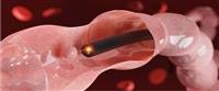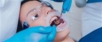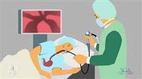
Gastroscopy or endoscopy is a medical procedure performed by a doctor, through which a tube called an endoscope is ed into the body and equipped with a camera and light at its end. The endoscope films a specific area inside the body, and sends images directly to the TV screen in front of the doctor.
The doctor moves the endoscope, and he can also use it to take a biopsy, a sample of cells or tissue for laboratory examination (Biopsy), and also to remove a polyp or to stop a blood vessel bleeding.
Upper gastrointestinal endoscopy includes
Esophagogastroduodenoscopy (Esophagus).
gastroscopy
Duodenum endoscopy (duodenum).
Since the duodenum is connected to the bile ducts and the pancreatic duct, this device can be used to perform a catheterization of these parts for the purpose of diagnosis and treatment. This is called ERCP- Endoscopic Retrograde Cholan Pancreatography. This catheter requires the injection of a contrast agent ) which improves the contrast and appearance of tissues.
This model can be used for examination and even treatment of bile ducts, such as: ing a stent into the bile duct to avoid blockage by a tumor.
In addition, an endoscope with an ultrasound transducer can be used to obtain information about diseases in or near the gastrointestinal tract. This type of endoscopic ultrasound is called EUS - Endoscopic Ultrasound.
When is the test performed?
Gastrointestinal endoscopy can be examined according to a pre-scheduled appointment or urgently, which is often in hospitals. The cases in which the test is performed include the following:
Check for complaints of upper abdominal or chest pain, heartburn, suspected stomach ulcers, or gastroesophageal reflux disease (GERD).
Check for complaints about difficulty or pain when swallowing, nausea, or vomiting.
Within the framework of the procedures to ascertain the incidence of anemia.
Check complaints of bloody vomiting, especially in patients with liver disease.
Esophageal varice treatment.
Diagnostics and treatment of diseases involving the bile ducts such as
Gallstones in the gallbladder.
Gallbladder catheterization (ERCP).
Evaluation of the extent of tumors spreading in the wall of the esophagus, stomach, pancreas, duodenum or around the pancreas and bile ducts with endoscopic ultrasound (EUS: Endoscopic Ultrasound).
The at-risk category
Each GI endoscopy test carries some minor risk of bleeding or perforation. This risk is very small, less than 0.1% with diagnostic endoscopy.
When endoscopy is used to also carry out therapeutic activities, such as: removing a tumor, the risk of this complication rises to 2%, for example: in the examination of the gallbladder catheterization there are additional risks ranging from mild inflammation of the pancreas to various infectious diseases and bleeding.
It is important to note that in many cases complications can be treated immediately, but sometimes surgery is required.
Related diseases
Diseases related to this examination include the following
Ulcers.
Gastroesophageal reflux disease (GERD).
Esophageal varices.
Tumors of the esophagus, stomach, duodenum, liver and pancreas.
bleeding
Mallory Weiss Tear Syndrome.
Gall stones.
How to perform the examination
You will be asked to remove your dentures from your mouth, and you will also be given an intravenous (IV) tube through which you will be given fluids and medications.
You are given numbing medication, then a member of the medical team places a rigid frame in your mouth. The goal of the frame is to prevent you from biting the speculum and injuring yourself.
Prepare for examination
Before examining the GI endoscopy, you must tell the doctor if you have a problem with one of the heart valves, that you need to take antibiotics before dental treatment or surgeries, if you have an artificial valve, or if you have suffered in the past from an infection in the heart valve called endocarditis ( Endocarditis).
It is forbidden to eat or drink 8 hours before the endoscopy of the gastrointestinal tract, and in the event of taking medications, the attending physician should be consulted.
During the examination
You will be asked to lie on your left side, while the doctor, who is often a gastroenterologist, will the endoscope into your mouth. event details.
The examination usually takes about 20 minutes, but may be longer when catheterization of the bile ducts or when performing interventional procedures.
after examination
You will be taken to the recovery room for 30 minutes, you may feel a burning sensation in your throat but it is transient, if everything is OK you are allowed to go home after a short time, or you may have to stay in the hospital to monitor your situation depending on your situation
Symptoms to watch out for after gastroscopy
Strong pain in the abdomen or chest.
Vomiting with or without blood.
Constant coughing.
Fever above 37.7 degrees Celsius.
Chills.
If one or more of these symptoms appear, you should refer to the doctor as soon as possible.
Warnings
During pregnancy
The doctor should be informed if there is a pregnancy.
Driving
The test is often done under sedation or superficial sedation, so you cannot drive after the test, and if you are not required to stay in hospital after the test, you should arrange for an escort to drive you home. Do not operate dangerous machinery or heavy equipment during the test. 8 hours after the test.
Medicines that affect the result of the examination
You must inform the doctor if you are allergic to the drug, and if you suffer from diseases of the heart or lungs, in case you have diabetes (diabetes), consult your doctor about the levels of diabetes the day before the examination.
It is important to consult your doctor, especially if you are taking warfarin-type blood-thinning medications.
Analysis of the results
After examining the gastrointestinal endoscopy, the doctor discusses the results with the patient. You may receive a page with details or a drawing of the results. In addition, colored pictures of the results may be attached. They may tell the patient that they have taken a biopsy, which is a sample of cells for laboratory examination, and the result should be awaited during Several days and up to 2-3 weeks.
A proper examination may contain the following elements
The mucosa is normal (Mucosa), without evidence of ulcers, inflammation, or tumors.
The course of the esophagus is normal, without narrowing.
The bile ducts are completely healthy without defects in function.