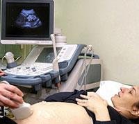
Ultrasound is the imaging method used today in pregnant women. Ultrasound imaging can be performed when pregnant through the abdomen or vagina, and it is the preferred method in the second and third trimesters of pregnancy. More details follow: 1. Ultrasound in the first trimester of pregnancy
In the first trimester of pregnancy, ultrasound is a tool that helps
Determine the gestational age.
Representation of the fetal heartbeat.
Locating pregnancy.
Determine the number of gestational sacs in a multi-embryo pregnancy.
In the first trimester, cervical labia examination and early instrumental scans are performed with the help of ultrasound imaging.
The cervical translucency examination is a measurement of the transparent area in the neck of the fetus, and it is the most predictive of Down syndrome, and is of great importance in the early diagnosis of cardiac abnormalities and a long list of other syndromes.
As for the early instrumental scan, it is performed in the 13-16th week of pregnancy, and its advantage lies in enabling the early diagnosis of structural abnormalities in the fetus, such as:
A deficiency in the development of the fetal brain.
Cardiac abnormalities.
Suspicious signs of genetic syndromes.
2. Ultrasound imaging in the second and third trimesters of pregnancy
In the second and third trimesters of pregnancy, an ultrasound follow-up is performed for the following reasons:
Dimensions of the fetus.
placenta site.
Data that represent his good condition, such as the amount of water and its movement.
In the second trimester of pregnancy in weeks 20-24, a late scan of the organs is performed, which contributes to the diagnosis of fetal deformities. There are known signs in ultrasound imaging of genetic syndromes such as Down syndrome, which can be detected in the early scan, as some of them disappear with time and cannot be detected. in the later survey.
When signs that raise suspicion of a specific syndrome are diagnosed with an ultrasound test, the woman is directed to receive genetic counseling.
Ultrasound imaging of the fetus
3. Ultrasound examination and invasive procedures
Ultrasound examination is a necessary tool for conducting invasive procedures with the aim of prenatal genetic diagnosis and with the aim of treating the fetus in life-threatening conditions in the womb, such as: Chorionic Villus Sampling-CVS.
It is performed in the 11-12 weeks of pregnancy through the vagina or through the abdomen by means of a needle, which is ed into the placental region with the aim of withdrawing the placental villi.
With the help of the placental villi examination, information related to fetal genes and hereditary diseases can be obtained, most notably the following:
Cystic fibrosis.
Hemophilia.
Fragile X Syndrome.
Tay-Sachs disease and others.
Amniocentesis is performed starting from the 16th week of pregnancy through the abdomen, as an amniocentesis is performed and a little fluid is withdrawn on which the above-mentioned tests can be performed.
In the past, the amniotic fluid was also examined to monitor the incompatibility of the blood type between the mother and the fetus, but today this examination has been replaced by non-invasive ultrasound imaging.
The reported rate of miscarriage after the two procedures is approximately 1%, while in multiple-fetal pregnancies, the rate of miscarriage is approximately 5%.