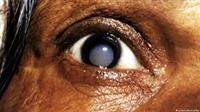

Keratoconus may occur in all age groups, so what are the symptoms of keratoconus? And what are its reasons?
Keratoconus is a pathological condition that affects the cornea of the eye, and it occurs when the cornea takes an abnormal shape, increases its curvature, and protrudes outward to transform from the spherical shape to the conical shape, due to the high pressure inside the eye.
Keratoconus often begins in the teens or twenties, but it can be diagnosed in childhood, and the shape of the cornea changes over a long period of time that may reach several years, but it develops faster in younger patients. Know the symptoms of keratoconus:
Symptoms of keratoconus
Keratoconus causes symptoms, including
Gradual loss of vision in one or both eyes, often in late adolescence.
Double vision when looking with one eye, even with glasses.
Seeing halos around bright lights.
Glare and sensitivity to bright light, making it difficult to drive at night.
Blurred vision and gradual deflection.
Sudden changes in vision in one eye.
Myopia, astigmatism.
And you should go to the ophthalmologist when you feel the rapid development of eye problems.
Causes of keratoconus
The cause of keratoconus is unknown, but genetic and environmental factors may contribute to the infection.
Risk factors for keratoconus
There are factors that increase the risk of infection and symptoms of keratoconus, including:
Having a family history of keratoconus.
Rubbing the eye vigorously.
Some medical conditions, such as: retinitis pigmentosa, Ehlers-Danlos syndrome, Down syndrome, hay fever, and asthma.
Diagnosis of keratoconus
To diagnose keratoconus symptoms, the degree of corneal curvature must be measured, and this is done through the following tests:
Ocular refraction
In which vision is checked by looking at a device that contains a wheel that contains several lenses, to determine which lens or combination of lenses gives the best vision to the patient.
Some doctors may use a hand-held instrument called a retinoscope to examine the eye.
Slit-lamp examination
By shining a beam of light perpendicular to the surface of the eye, and examining the eye through a microscope to determine the shape of the cornea and to look for any other problems.
Keratometry
It is used to determine the shape of the cornea by shining a circle of light on the cornea and measuring the degree of reflection.
Computerized keratoplasty
Pictures of the cornea are recorded by examining the imaging of optical correlation and topography of the cornea to make a colorful and detailed map of the surface of the cornea, and to determine the thickness of the cornea.