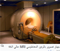
It is a medical imaging method that relies on X-rays (X-rays) used to a three-dimensional image of the internal organs of the body. It consists of several two-dimensional images taken about a fixed axis of rotation.
Computerized tomography has a very high image clarity and shows bone details very precisely, unlike magnetic resonance imaging, which depicts soft tissues with high accuracy, and this procedure is pain-free.
The information and images resulting from the tomography device are processed to show the body’s organs according to their ability to prevent the passage of x-rays through them, and also this information can be used to draw three-dimensional images, and the images obtained are cross-sectional in nature, and each two-dimensional imaging slice resembles, for example, what You see it when you cut an apple in half through the middle and then look at the surface of the cut If you start from the top of the apple down to the bottom you will get a series of slices that represent the overall structure. With the help of a computer, three-dimensional images can be reconstructed from these two-dimensional slices. The use of this type of device is very common around the world, for example, in the United States 80 million people undergo this type of imaging annually, and 50 million in Japan.
Scientists fear that using this type of imaging in excess may lead to cancers. Doctors are not the only ones who use this device, archaeologists also use it to examine ancient artifacts such as coffins. It is called because this method gives x-ray images in the form of sections of the body, computed tomography is carried out using a special device called a computed tomography device or computed tomography scanner, This method is characterized by its accuracy, gives clear images, and can give images of places that may be difficult to photograph with conventional radiography, as well as it can be done quickly and accurately.
Uses
Computerized tomography is used to diagnose many diseases such as malignant and benign tumors in different areas of the body.
the brain
CT scanning of the head is used to diagnose tumors, internal bleeding, and calcifications. Tumors are easily noticeable because they swell and increase in size, causing damage to nearby tissues. Ambulances are often equipped with small CT scanners to screen people who have had a stroke or a blow to the head. A special type of this machine called (N-localizer) is often used in surgical operations performed when extirpating an intracranial tumor or treating arteriovenous malformations. The brain can also be scanned by an MRI machine, but some diseases and tumors can only be observed through Computed tomography.
the chest
Computerized tomography is used to diagnose and observe changes in the spongy inner lung tissue. This type of change cannot be observed with plain X-rays and MRIs. In addition, this scan is used to find cancerous and other tumors in the lungs. Often a CT scan is taken twice, once on the inhale and once on the exhale, this process is called a high-resolution CT scan. Patients usually undergo many tests within a short period of time using these devices to monitor changes in abnormal things in their chest, such as tumors and infections. This is very harmful because excessive exposure to X-rays can cause damage to the body.
This particular type of image has been used to diagnose, confirm or deny a person''s infection with the novel coronavirus.
The heart (important in the diagnosis of coronary artery, calcification rate with injury to three arteries)
abdomen and pelvis
It is used in arteriovenous imaging instead of interventional radiology, which is safer, takes a short time, and diagnoses pulmonary embolism instead of nuclear imaging.
Advantages of computed tomography
There are many characteristics that make this method of surveying better than the rest of the medical survey methods. First, tomography can show a clear picture of the organ being imaged without showing the organs that surround it. For example, when imaging the lungs, the heart or viscera do not appear in the image. Second, the color contrast between the tissues in the image helps doctors tell the difference in mass density. In addition, this mechanism can produce high-quality images without emitting a large amount of radiation. The most important feature of this mechanism is that no gas or device has to be placed directly inside the body, as is the case with a catheter.