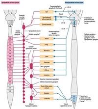
The sympathetic nervous system is part of the autonomic nervous system, along with its counterpart, parasympathetic nervous system.
The origin of the sympathetic nervous system is found within the thoracic and lumbar segments of the spinal cord also known as the thoracolumbar division (T1 to L2,3).
The sympathetic pathway can be divided into three following components
The preganglionic neurons,
The sympathetic ganglia,
The postganglionic neurons.
While the sympathetic nervous system is also important at rest, it is essential for preparing the body for emergency response in endangering situations, also known as the “fight-or-flight” response. The sympathetic system activates numerous complex pathways to enable an adequate response to a threat or trauma. Some of these physical effects include faster breathing, increased heart rate and blood pressure, dilation of pupils, redirection of blood flow to important organs (e.g. brain and muscles), and increased sweating.
This article will discuss the anatomy and function of the sympathetic nervous system.
Autonomic (visceral) nervous system
The nervous system can be divided into central and peripheral nervous systems. The peripheral nervous system is further divided into the somatic and autonomic nervous systems.
The autonomic nervous system (ANS) is a functional division of the nervous system that controls involuntary actions of muscles, glands and internal organs (e.g. bowel movements). Together with endocrine glands, the ANS affects important body functions without the direct involvement of the cerebral cortex. In contrast, the somatic nervous system mediates voluntary responses of the body (e.g. skeletal muscle function) and it’s under the direct control of the cerebral cortex.
The ANS can be divided according to its location (central and peripheral parts) and function. Functionally, the ANS is divided into sympathetic (SNS) and parasympathetic (PSNS) nervous systems. They usually work antagonistically in the organs but in a well-integrated manner. It is the balance of the actions of both divisions that maintains a stable internal environment in the body.
The anatomical distinction between the sympathetic and parasympathetic divisions is given by the location of the presynaptic cell bodies and the types of nerves conducting presynaptic nerve fibers.
General sympathetic pathway
The general sympathetic pathway can be simplified into the following components
Preganglionic neurons
Sympathetic ganglia
Postganglionic neurons.
The preganglionic neurons located inside the thoracic and lumbar segments (T1-L2,3) of the spinal cord and their fibers (axons), which are called preganglionic fibers. These preganglionic fibers synapse with the postganglionic neurons inside the sympathetic ganglia, which are typically found near the vertebral column. These ganglia are actually a collection of cell bodies of postganglionic neurons. The postganglionic neurons give off long axons (postganglionic fibers) that leave the ganglia and project onto visceral effectors, where they release the neurotransmitter norepinephrine. Both preganglionic and postganglionic neurons are multipolar.
Preganglionic neurons
The cell bodies of the preganglionic neurons of the SNS are found only in the intermediolateral cell columns (ICLs) of the spinal cord, one on the left side and on the right. ICLs are part of the lateral horns of the gray matter of the thoracic (T1-12) and upper lumbar (L2 or L3) spinal cord segments, hence the native name “thoracolumbar” for the sympathetic division. This region consists of the visceral motor region of the spinal gray matter. You can think of the ICLs as longitudinal tubes passing through the respective lateral horns of the spinal cord. The preganglionic SNS cell bodies are organised somatotopically, meaning the arrangement of the cell bodies is a close representation to that of the body. Basically, the T1-6 cell bodies that are located superiorly innervate the head, upper limb and thoracic viscera. T7-11 located in the middle innervate the body wall and abdominal viscera, while T11-L2(3) located inferiorly innervate the lower limb and pelvic viscera.
The preganglionic fibers leave the ICLs and thus, the spinal cord through the anterior roots. They travel very briefly through the anterior rami of spinal nerves T1-L2(3), before leaving them and passing to the sympathetic trunks (more details later) through the white rami communicantes (white because nerve fibers are covered with white myelin).
Sympathetic ganglia
The ganglionic compartment is actually composed of the cell bodies of the postganglionic neurons. It consists of two types, paravertebral and prevertebral ganglia.
Paravertebral ganglia (“para” = alongside, beside) occur on either side of the vertebral column and are independently linked on either side, forming two sympathetic trunks (chains). The paravertebral ganglia are the site where preganglionic fibers synapse with postganglionic neurons. The trunks extend the entire length of the column, from the base of the cranium to the coccyx. They converge anteriorly at the coccyx, forming the ganglion impar (ganglion of Walther). Each trunk is attached to the anterior rami of the T1-L2(3) spinal nerves.
Prevertebral ganglia (splanchnic ganglia) are located in the abdominal cavity around the origin of the major branches of the abdominal aorta. The prevertebral ganglia form aggregations around the abdominal prevertebral plexus and are referred to as the celiac, aorticorenal and superior and inferior mesenteric ganglia. Various nerve plexuses branch from these ganglia.
Course of fibers
In general, after passing briefly through the anterior rami, preganglionic fibers enter the sympathetic trunk via white rami communicantes. Inside the trunk, preganglionic fibers can follow one of four courses:
1. Ascend and synapse in a higher paravertebral ganglion
Within the sympathetic trunk, preganglionic fibers usually from T1-5 spinal cord levels can ascend to other vertebral levels and synapse inside ganglia located at a more superior level. The ganglia might not necessarily be associated with inputs directly from the spinal cord (other nerves than T1-L2/3 can participate in the synapse).
2. Descend and synapse in a lower paravertebral ganglion
These are similar to the ascending preganglionic, but in contrast, they descend to ganglia located at a more inferior level. This pathway usually involves fibers from T5-L2(3). The ascending and descending preganglionic fibers gives the sympathetic trunk the appearance of a chain with connections between the ganglia.