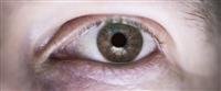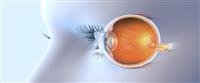

Retinal detachment is a serious condition that can lead to complete loss of vision if not treated directly. Retinal detachment disease The retina is one of the sensitive and important areas in the human body as it is responsible for sending visual signals through the optic nerve to the brain, which in turn translates them into the image we see. A serious condition because it may cause total blindness if not treated as quickly as possible.
Retinal detachment occurs when the inner lining of the eye (the retina) separates from the eye wall (the placenta), which contains blood vessels that supply the eye with oxygen and necessary nutrients.
Types of retinal detachment
There are 3 different types of retinal detachment
Rhegmatogenous retinal detachment: It occurs as a result of holes or gaps in the retina, which in turn allows fluid to pass under the retina and separates it from the placenta as a result of this rupture.
Exudative retinal detachment: This is an inflammatory disorder that causes fluid to build up behind the retina.
Tractional retinal detachment: It occurs when fibrous tissue forms that stretches the retina, creating holes in it.
Symptoms of retinal detachment
Here are the main symptoms that indicate a possible retinal detachment
Sudden appearance of black dots, spots, or lines floating within the field of vision.
Seeing a large number of floating objects, some may notice one large black shape floating within the field of vision like a fly.
Suffering from sudden short flashes of light, not exceeding one second, that appear in front of the affected eye.
Seeing a dark shadow that appears as a curtain from the outer side of the field of vision in the affected eye.
Sometimes blurred vision.
Causes of retinal detachment
Are you at risk of retinal detachment? The answer is yes in the cases below
Advancing age. Retinal detachment is more common in people over the age of 50.
Previous occurrence of retinal detachment in one eye.
Having a family history of retinal detachment.
Extreme nearsightedness.
Previous eye surgery, such as cataract removal.
Exposure to disease or inflammation in the other eye before.
Suffering from diabetes, as diabetics are more susceptible to retinal detachment.
Diagnosis of retinal detachment
The doctor may use the following tests to diagnose retinal detachment
A normal vision test to measure your vision and ability to distinguish colors.
Examination of the retina by using an instrument equipped with a bright light and special lenses to examine the back of the eye, including the retina.
Ultrasound imaging is used if bleeding occurs in the eye, making it difficult to see the retina.
Examination of intraocular pressure and blood flow throughout your eye, specifically in the retina.
The doctor tests the retina''s ability to send nerve signals to your brain.
Retinal detachment treatment
It is necessary to treat retinal detachment, because one of the important consequences of neglecting retinal detachment treatment is irreversible vision loss due to retinal atrophy or chronic optic inflammation, which makes the entire eye vulnerable to atrophy.
Surgical treatment is always the choice in order to repair tears and holes that cause retinal detachment by means of a laser or a cold retinal probe to return the retina to its position, as it works to push the back wall of the eye from the outside towards the retina.
This is done by suspending strips or parts of silicone or removing the vitreous and ing silicone gas or oil into the vitreous cavity. The operation is done under local anesthesia and does not require the patient to be kept in the hospital.