

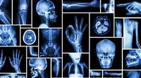
X-rays are typically done first. They are most valuable for detecting abnormalities in bone and are taken to evaluate painful, deformed, or suspected abnormal areas of bone. Often, x-rays can help to diagnose fractures, tumors, injuries, infections, and deformities (such as developmental dysplasia of the hip). Also, sometimes x-rays are helpful in showing changes that confirm a person has a certain kind of arthritis (for example, rheumatoid arthritis or osteoarthritis). X-rays do not show soft tissues such as muscles, bursae, ligaments, tendons, or nerves.
To help determine whether the joint has been damaged by injury, a doctor may use an ordinary (non-stress) x-ray or one taken with the joint under stress caused by certain positions (stress x-ray).
Arthrography is an x-ray procedure in which a radiopaque dye is injected into a joint space to outline the structures, such as ligaments inside the joint. Arthrography can be used to view torn ligaments and fragmented cartilage in the joint. However, magnetic resonance imaging (MRI) is now generally used in preference to arthrography.


